Snapp Erik Lee, Lajoie Patrick
Cold Spring Harb Protoc. 2011 Nov 1;2011(11):1295-304. doi: 10.1101/pdb.top066548.
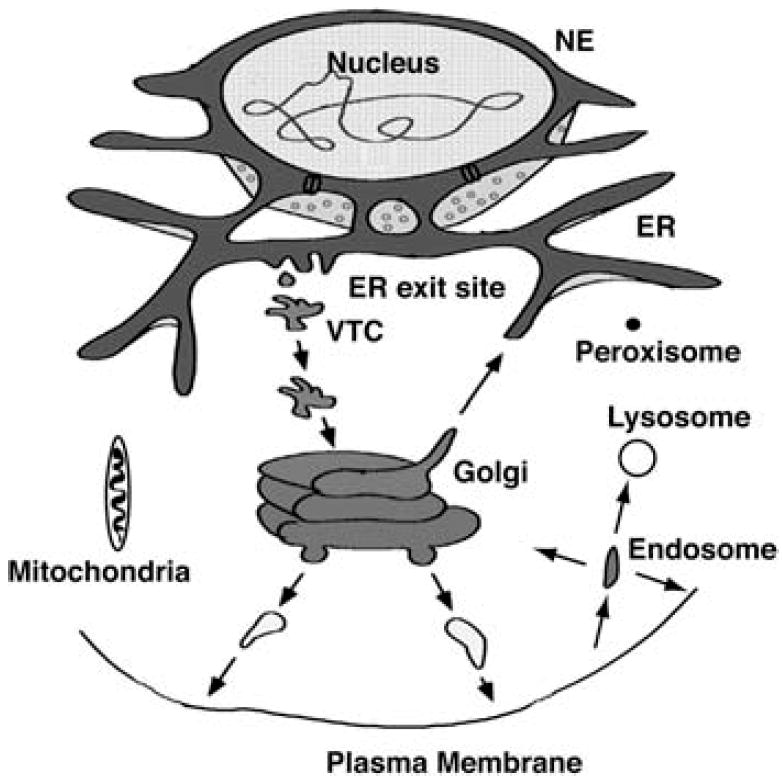
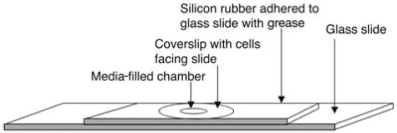
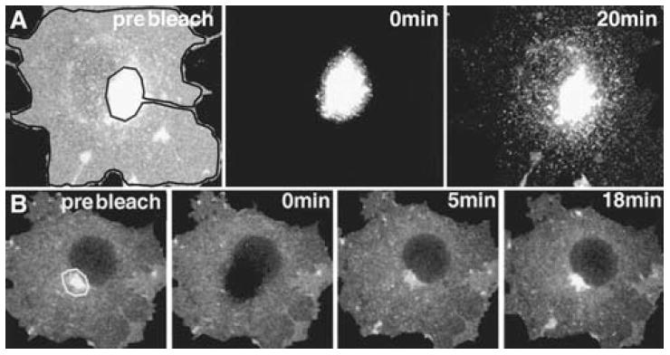
Eukaryotic cells are composed of an intricate system of internal membranes that are organized into different compartments--including the endoplasmic reticulum (ER), the nuclear envelope, the Golgi complex (GC), lysosomes, endosomes, caveolae, mitochondria, and peroxisomes--that perform specialized tasks within the cell. The localization and dynamics of intracellular compartments are now being studied in living cells because of the availability of green fluorescent protein (GFP)-fusion proteins and recent advances in fluorescent microscope imaging systems. Results using these techniques are revealing how intracellular compartments maintain their steady-state organization and distributions, how they undergo growth and division, and how they transfer protein and lipid components between themselves through the formation and trafficking of membrane transport intermediates. This article describes methods using GFP-fusion proteins to visualize the behavior of organelles and to track membrane-bound transport intermediates moving between them. Practical issues related to the construction and expression of GFP-fusion proteins are discussed first. These are essential for optimizing the brightness and expression levels of GFP-fusion proteins so that intracellular membrane-bound structures containing these fusion proteins can be readily visualized. Next, techniques for performing time-lapse imaging using a confocal laser-scanning microscope (CLSM) are detailed, including the use of photobleaching to highlight organelles and transport intermediates. Methods for the acquisition and analysis of data are then discussed. Finally, commonly used and exciting new approaches for perturbing membrane traffic are outlined.
真核细胞由一个复杂的内膜系统组成,该系统被组织成不同的区室,包括内质网(ER)、核膜、高尔基体复合体(GC)、溶酶体、内体、小窝、线粒体和过氧化物酶体,它们在细胞内执行特定任务。由于绿色荧光蛋白(GFP)融合蛋白的可用性以及荧光显微镜成像系统的最新进展,细胞内区室的定位和动态现在正在活细胞中进行研究。使用这些技术的结果正在揭示细胞内区室如何维持其稳态组织和分布,它们如何进行生长和分裂,以及它们如何通过膜转运中间体的形成和运输在彼此之间转移蛋白质和脂质成分。本文介绍了使用GFP融合蛋白来可视化细胞器行为并追踪在它们之间移动的膜结合转运中间体的方法。首先讨论与GFP融合蛋白的构建和表达相关的实际问题。这些对于优化GFP融合蛋白的亮度和表达水平至关重要,以便能够轻松可视化包含这些融合蛋白的细胞内膜结合结构。接下来详细介绍使用共聚焦激光扫描显微镜(CLSM)进行延时成像的技术,包括使用光漂白来突出细胞器和转运中间体。然后讨论数据采集和分析的方法。最后概述了用于干扰膜运输的常用和令人兴奋的新方法。