Harrill Joshua A, Chen Hao, Streifel Karin M, Yang Dongren, Mundy William R, Lein Pamela J
Mol Brain. 2015 Feb 15;8:10. doi: 10.1186/s13041-015-0099-9.
Synaptogenesis is a critical neurodevelopmental process whereby pre- and postsynaptic neurons form apposed sites of contact specialized for chemical neurotransmission. Many neurodevelopmental disorders are thought to reflect altered patterns of synaptic connectivity, including imbalances between excitatory and inhibitory synapses. Developing rapid throughput approaches for assessing synaptogenesis will facilitate toxicologic and drug screening studies of neurodevelopmental disorders. The current study describes the use of high-content imaging to quantify the ontogeny of excitatory and inhibitory synapses using in vitro models of neurodevelopment. These data are compared to biochemical and functional measures of synaptogenesis.
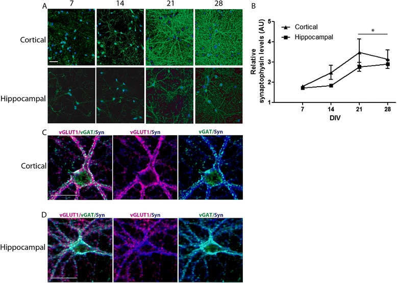
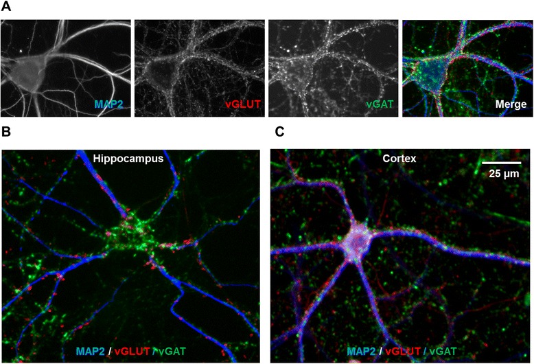
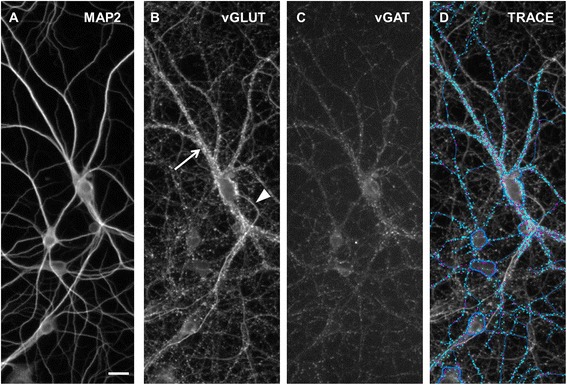
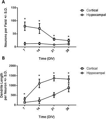
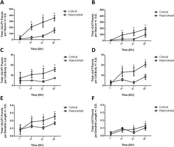
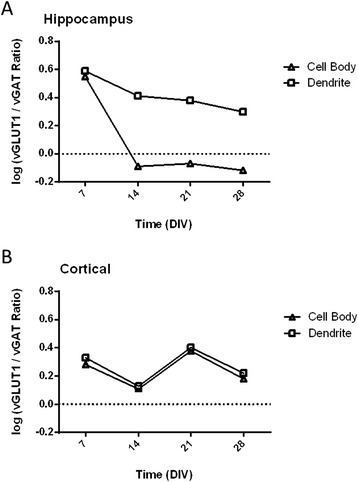
The ontogenetic patterns of synapse formation were compared between primary rodent hippocampal and cortical neurons over 28 days in vitro (DIV). As determined by ELISA, the increase in synaptophysin expression levels as cultures matured was similar between hippocampal and cortical cultures. High-content imaging of immunoreactivity of excitatory and inhibitory synaptic biomarkers demonstrated an overall greater number of synapses in hippocampal relative to cortical neurons with marked differences in the pattern of inhibitory synapse development between these two neuronal cell types. Functional assays revealed that both the mean firing rates and mean bursting rates were significantly increased in cortical cultures relative to hippocampal cultures. This difference may reflect decreased inhibitory synaptic tone in cortical versus hippocampal cultures.
These data demonstrate differences and similarities in the ontogeny of synaptogenesis between hippocampal and cortical neurons, depending on the biological level examined. Assessment of synaptophysin protein levels by ELISA showed a general increase in synapse formation in both cell types with increasing time in culture, while high-content imaging was able to delineate cell type-dependent differences in formation of excitatory versus inhibitory synapses. The functional significance of differences in the balance of excitatory to inhibitory synapses was confirmed by the assessment of network activity using microelectrode arrays. These results suggest that high-content imaging and microelectrode arrays provide complementary approaches for quantitative assessment of synaptogenesis, which should provide a robust readout of toxicologic and pharmacologic effects on this critical neurodevelopmental event.
突触发生是一个关键的神经发育过程,在此过程中,突触前神经元和突触后神经元形成专门用于化学神经传递的对置接触位点。许多神经发育障碍被认为反映了突触连接模式的改变,包括兴奋性和抑制性突触之间的失衡。开发用于评估突触发生的快速高通量方法将有助于神经发育障碍的毒理学和药物筛选研究。当前的研究描述了使用高内涵成像通过神经发育的体外模型来量化兴奋性和抑制性突触的个体发育。将这些数据与突触发生的生化和功能测量结果进行比较。
在体外培养28天(DIV)期间,比较了原代啮齿动物海马神经元和皮层神经元之间突触形成的个体发育模式。通过酶联免疫吸附测定(ELISA)确定,随着培养物成熟,海马和皮层培养物中突触素表达水平的增加相似。对兴奋性和抑制性突触生物标志物免疫反应性的高内涵成像显示,相对于皮层神经元,海马中的突触总数总体上更多,并且这两种神经元细胞类型之间抑制性突触发育模式存在明显差异。功能测定表明,相对于海马培养物,皮层培养物中的平均放电率和平均爆发率均显著增加。这种差异可能反映了皮层培养物与海马培养物相比抑制性突触张力降低。
这些数据表明,根据所检查的生物学水平,海马神经元和皮层神经元在突触发生个体发育方面存在差异和相似之处。通过ELISA评估突触素蛋白水平显示,随着培养时间的增加,两种细胞类型中的突触形成普遍增加,而高内涵成像能够描绘出兴奋性和抑制性突触形成中细胞类型依赖性差异。使用微电极阵列评估网络活动证实了兴奋性与抑制性突触平衡差异的功能意义。这些结果表明,高内涵成像和微电极阵列为突触发生的定量评估提供了互补方法,这应该能够有力地读出对这一关键神经发育事件的毒理学和药理学影响。