Calvieri Camilla, Masselli Gabriele, Monti Riccardo, Spreca Matteo, Gualdi Gian Franco, Fedele Francesco
Department of Cardiovascular, Respiratory, Nephrologic and Geriatric Sciences, La Sapienza University of Rome, Viale del Policlinico 155, 00161 Rome, Italy.
Department of Radiology, La Sapienza University of Rome, Viale del Policlinico 155, 00161 Rome, Italy.
Biomed Res Int. 2015;2015:859073. doi: 10.1155/2015/859073. Epub 2015 Feb 1.
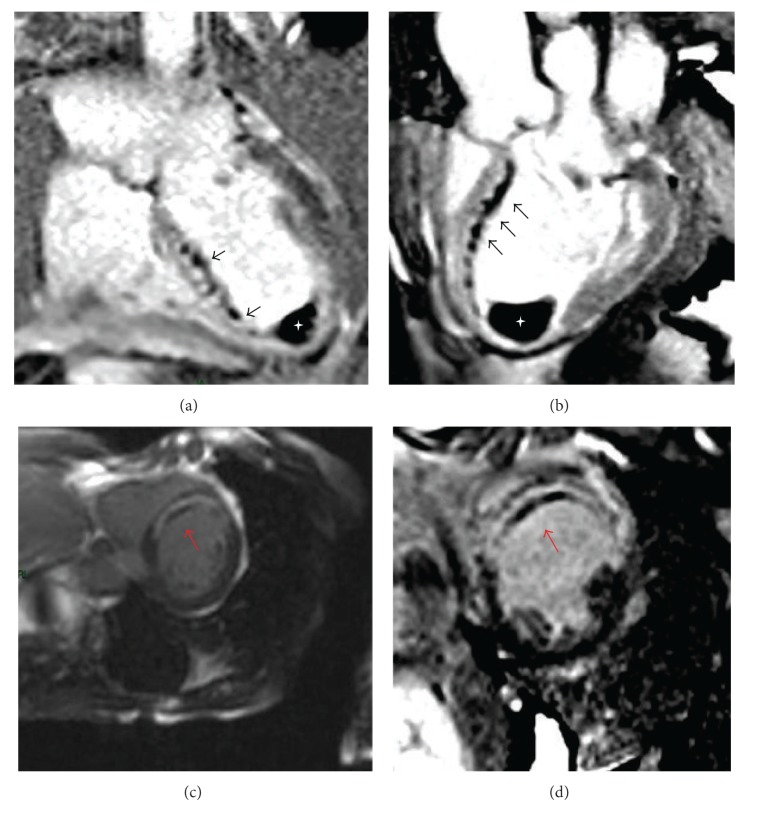
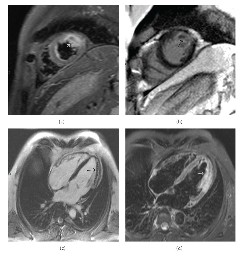
Cardiovascular magnetic resonance (CMR) is a useful noninvasive technique for determining the presence of microvascular obstruction (MVO) and intramyocardial hemorrhage (IMH), frequently occurring in patients after reperfused myocardial infarction (MI). MVO, or the so-called no-reflow phenomenon, is associated with adverse ventricular remodeling and a poor prognosis during follow-up. Similarly, IMH is considered a severe damage after revascularization by percutaneous primary coronary intervention (PPCI) or fibrinolysis, which represents a worse prognosis. However, the pathophysiology of IMH is not fully understood and imaging modalities might help to better understand that phenomenon. While, during the past decade, several studies examined the distribution patterns of late gadolinium enhancement with different CMR sequences, the standardized CMR protocol for assessment of IMH is not yet well established. The aim of this review is to evaluate the available literature on this issue, with particular regard to CMR sequences. New techniques, such as positron emission tomography/magnetic resonance imaging (PET/MRI), could be useful tools to explore molecular mechanisms of the myocardial infarction healing process.
心血管磁共振成像(CMR)是一种有用的非侵入性技术,可用于确定微血管阻塞(MVO)和心肌内出血(IMH)的存在,这在再灌注心肌梗死(MI)患者中经常出现。MVO,即所谓的无复流现象,与不良的心室重构和随访期间的不良预后相关。同样,IMH被认为是经皮冠状动脉介入治疗(PPCI)或纤维蛋白溶解血管重建后的严重损伤,这代表着更差的预后。然而,IMH的病理生理学尚未完全了解,成像方式可能有助于更好地理解这一现象。在过去十年中,虽然有几项研究使用不同的CMR序列检查了钆延迟强化的分布模式,但用于评估IMH的标准化CMR方案尚未完全确立。本综述的目的是评估关于这个问题的现有文献,特别是关于CMR序列的文献。新技术,如正电子发射断层扫描/磁共振成像(PET/MRI),可能是探索心肌梗死愈合过程分子机制的有用工具。