• 文献检索
  • 文档翻译
  • 深度研究
  • 学术资讯
  • Suppr Zotero 插件Zotero 插件
  • 邀请有礼
  • 套餐&价格
  • 历史记录
应用&插件
Suppr Zotero 插件Zotero 插件浏览器插件Mac 客户端Windows 客户端微信小程序
定价
高级版会员购买积分包购买API积分包
服务
文献检索文档翻译深度研究API 文档MCP 服务
关于我们
关于 Suppr公司介绍联系我们用户协议隐私条款
关注我们

Suppr 超能文献

核心技术专利:CN118964589B侵权必究
粤ICP备2023148730 号-1Suppr @ 2026

文献检索

告别复杂PubMed语法,用中文像聊天一样搜索,搜遍4000万医学文献。AI智能推荐,让科研检索更轻松。

立即免费搜索

文件翻译

保留排版,准确专业,支持PDF/Word/PPT等文件格式,支持 12+语言互译。

免费翻译文档

深度研究

AI帮你快速写综述,25分钟生成高质量综述,智能提取关键信息,辅助科研写作。

立即免费体验

隆鼻患者锥形束CT中窦口鼻道复合体的解剖变异

Anatomical Variations of Ostiomeatal Complex in CBCT of Patients Seeking Rhinoplasty.

作者信息

Khojastepour Leila, Mirhadi Sabah, Mesbahi Seyed Alireza

机构信息

Dept. of Oral and Maxillofacial Radiology, School of Dentistry, Shiraz University of Medical Science, Shiraz, Iran.

Dept. of Otolaryngology and Plastic Surgery, Khodadoust Hospital, Shiraz Iran.

出版信息

J Dent (Shiraz). 2015 Mar;16(1):42-8.

PMID:25759857
原文链接:https://pmc.ncbi.nlm.nih.gov/articles/PMC4345113/
Abstract

STATEMENT OF THE PROBLEM

Anatomic variation can potentially impact the surgical safety.

PURPOSE

The purpose of this cross-sectional study was to assess the prevalence of ostiomeatal complex variations based on cone beam computed tomography (CBCT) images of the patients seeking rhinoplasty.

MATERIALS AND METHOD

In this cross-sectional study, CBCT images of 281 patients including 153 female and 128 male with Mean±SD age of 26.97±7.38 were retrieved and analyzed for presence of variations of ostiomeatal complex and mucosal thickening. All CBCT images were acquired by NewTom VGi scanner with 15×15 field of view, as a part of preoperative recording of patients seeking rhinoplasty in an otolaryngology clinic. Chi- square test and Odds ratio were used for statistical analysis of the obtained data and p< 0.05 was considered to be statistically significant.

RESULTS

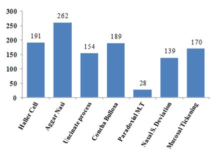
Agger nasi cells which were seen in 93.2% of the cases were the most common anatomic variation. It was followed by Haller cells (68%), concha bullosa (67.3%), uncinate process variations (54.8%), nasal sepal deviation (49.5%) and paradoxical curvature of middle turbinate (10%). Mucosal thickening were detected in 60.7% of the studied cases.

CONCLUSION

Ostiomeatal complex variations and mucosal thickening are considerably prevalent among the patients seeking rhinoplasty. This study also revealed that CBCT evaluation of paranasal sinuses has comparable result in delineation of the sinonasal anatomy.

摘要

问题陈述

解剖变异可能会影响手术安全性。

目的

本横断面研究的目的是基于接受隆鼻手术患者的锥形束计算机断层扫描(CBCT)图像评估窦口鼻道复合体变异的患病率。

材料与方法

在这项横断面研究中,检索并分析了281例患者的CBCT图像,其中包括153名女性和128名男性,平均年龄±标准差为26.97±7.38岁,以确定是否存在窦口鼻道复合体变异和黏膜增厚。所有CBCT图像均由视野为15×15的NewTom VGi扫描仪采集,作为一家耳鼻喉科诊所接受隆鼻手术患者术前记录的一部分。采用卡方检验和比值比对所得数据进行统计分析,p<0.05被认为具有统计学意义。

结果

在93.2%的病例中可见鼻丘气房,是最常见的解剖变异。其次是Haller气房(68%)、泡状鼻甲(67.3%)、钩突变异(54.8%)、鼻中隔偏曲(49.5%)和中鼻甲反常弯曲(10%)。在60.7%的研究病例中检测到黏膜增厚。

结论

窦口鼻道复合体变异和黏膜增厚在接受隆鼻手术的患者中相当普遍。本研究还表明,CBCT对鼻窦的评估在描绘鼻窦解剖结构方面具有可比的结果。

https://cdn.ncbi.nlm.nih.gov/pmc/blobs/a5ef/4345113/58cb7eb05a10/jds-16-42-g002.jpg
https://cdn.ncbi.nlm.nih.gov/pmc/blobs/a5ef/4345113/155d25308ce8/jds-16-42-g001.jpg
https://cdn.ncbi.nlm.nih.gov/pmc/blobs/a5ef/4345113/58cb7eb05a10/jds-16-42-g002.jpg
https://cdn.ncbi.nlm.nih.gov/pmc/blobs/a5ef/4345113/155d25308ce8/jds-16-42-g001.jpg
https://cdn.ncbi.nlm.nih.gov/pmc/blobs/a5ef/4345113/58cb7eb05a10/jds-16-42-g002.jpg

相似文献

1
Anatomical Variations of Ostiomeatal Complex in CBCT of Patients Seeking Rhinoplasty.隆鼻患者锥形束CT中窦口鼻道复合体的解剖变异
J Dent (Shiraz). 2015 Mar;16(1):42-8.
2
A comparative study of paranasal sinus and nasal cavity anatomic variations between the Polish and Turkish Cypriot Population with CBCT.基于 CBCT 的波兰和土耳其塞浦路斯人群鼻窦和鼻腔解剖结构变异的对比研究。
Head Face Med. 2022 Nov 26;18(1):37. doi: 10.1186/s13005-022-00340-3.
3
Correlations between anatomical variations of the nasal cavity and ethmoidal sinuses on cone-beam computed tomography scans.锥形束计算机断层扫描鼻窦鼻腔解剖变异之间的相关性。
Imaging Sci Dent. 2019 Jun;49(2):103-113. doi: 10.5624/isd.2019.49.2.103. Epub 2019 Jun 24.
4
[Computerized tomography in the evaluation of anatomic variations of the ostiomeatal complex].[计算机断层扫描在评估窦口鼻道复合体解剖变异中的应用]
Radiol Med. 1993 Sep;86(3):195-9.
5
CT scan evaluation of the anatomical variations of the ostiomeatal complex.鼻窦开口复合体解剖变异的CT扫描评估
Indian J Otolaryngol Head Neck Surg. 2009 Sep;61(3):163-8. doi: 10.1007/s12070-009-0059-8. Epub 2009 Sep 27.
6
Assessing the prevalence of paranasal sinuses anatomical variants in patients with sinusitis using Cone Beam Computer Tomography.使用锥形束计算机断层扫描评估鼻窦炎患者鼻旁窦解剖变异的患病率。
Clujul Med. 2016;89(3):419-21. doi: 10.15386/cjmed-598. Epub 2016 Jul 28.
7
[Anatomic variations of the ostiomeatal complex and their correlation with chronic sinusitis: CT evaluation].[窦口鼻道复合体的解剖变异及其与慢性鼻窦炎的相关性:CT评估]
Zhonghua Er Bi Yan Hou Ke Za Zhi. 1999 Jun;34(3):143-6.
8
Agreement between cone-beam computed tomography and functional endoscopic sinus surgery for detection of pathologies and anatomical variations of the paranasal sinuses in chronic rhinosinusitis patients: A prospective study.锥形束计算机断层扫描与功能性鼻内镜鼻窦手术在慢性鼻窦炎患者鼻窦病变及解剖变异检测中的一致性:一项前瞻性研究。
Imaging Sci Dent. 2020 Dec;50(4):299-307. doi: 10.5624/isd.2020.50.4.299. Epub 2020 Dec 15.
9
Are of Osteomeatal Complex Variations Related to Nasolacrimal Canal Morphometry.筛窦骨管复合体变异与鼻泪管形态测量学的关系。
Iran J Otorhinolaryngol. 2022 Jan;34(120):17-26. doi: 10.22038/IJORL.2021.52612.2784.
10
Sinonasal Anatomical Variants: CT and Endoscopy Study and Its Correlation with Extent of Disease.鼻窦解剖变异:CT与内镜研究及其与疾病范围的相关性
Indian J Otolaryngol Head Neck Surg. 2016 Sep;68(3):352-8. doi: 10.1007/s12070-015-0920-x. Epub 2015 Sep 29.

引用本文的文献

1
Distribution and laterality of concha bullosa in patients with different cranial skeletal types: a retrospective analysis among cases with concha bullosa.不同颅骨骨骼类型患者中泡状鼻甲的分布及侧别:泡状鼻甲病例的回顾性分析
Maxillofac Plast Reconstr Surg. 2025 May 8;47(1):9. doi: 10.1186/s40902-025-00463-y.
2
Retrospective Study on the Prevalence of Haller Cells Using Cone Beam Computed Tomography.使用锥形束计算机断层扫描对哈勒氏细胞患病率的回顾性研究。
Cureus. 2024 Aug 19;16(8):e67200. doi: 10.7759/cureus.67200. eCollection 2024 Aug.
3
Comparison of Anthropometric and Cephalometric Measurements Obtained by Stereophotogrammetry and 3D Computed Tomography of the Nose Before Septorhinoplasty.

本文引用的文献

1
Clinically significant anatomical variants of the paranasal sinuses.鼻窦具有临床意义的解剖变异。
Oman Med J. 2014 Mar;29(2):110-3. doi: 10.5001/omj.2014.27.
2
Cone beam CT analysis of Haller cells: prevalence and clinical significance.Haller 细胞的锥形束 CT 分析:患病率和临床意义。
Dentomaxillofac Radiol. 2013;42(9):20130055. doi: 10.1259/dmfr.20130055. Epub 2013 Aug 23.
3
Rhinoplasty from a rhinologist's perspective: need for recognition of associated sinonasal conditions.从鼻科医生的角度看鼻整形术:需要认识到相关的鼻-鼻窦情况。
鼻中隔成形术前立体摄影和三维计算机断层扫描获得的鼻测量值的比较。
Aesthetic Plast Surg. 2024 Nov;48(21):4314-4322. doi: 10.1007/s00266-024-04097-9. Epub 2024 May 16.
4
[Applications of cone beam computed tomography of the craneofacial structure in medical specialties. A review].[锥形束计算机断层扫描在颌面结构医学专业中的应用。综述]
Rev Cient Odontol (Lima). 2022 Mar 30;10(1):e100. doi: 10.21142/2523-2754-1001-2022-100. eCollection 2022 Jan-Mar.
5
Evaluation of diagnostic accuracy of cone beam computed tomography and multi-detector computed tomography for detection of anatomical variations in rhinoplasty.评价锥形束 CT 和多排螺旋 CT 检测鼻整形术解剖变异的诊断准确性。
Head Face Med. 2024 Jan 3;20(1):1. doi: 10.1186/s13005-023-00401-1.
6
Cone Beam CT Analysis of Haller Cells: Prevalence and Relationship with Orbital Floor Dehiscence.锥形束CT对哈勒气房的分析:发生率及其与眶底裂开的关系
Int J Dent. 2023 Jan 31;2023:5200152. doi: 10.1155/2023/5200152. eCollection 2023.
7
Inter Observer Agreement Among Radiologist and Otorhinolaryngologists on Paranasal Sinus Computed Tomography Scans in Chronic Rhinosinusitis.放射科医生与耳鼻喉科医生对慢性鼻-鼻窦炎患者鼻窦计算机断层扫描结果的观察者间一致性研究
Indian J Otolaryngol Head Neck Surg. 2022 Dec;74(4):501-509. doi: 10.1007/s12070-021-03016-7. Epub 2022 Jan 7.
8
A Study of Anatomical Variations in the Nasal Cavity in Cases of Chronic Rhinosinusitis.慢性鼻窦炎病例鼻腔解剖变异的研究
Indian J Otolaryngol Head Neck Surg. 2022 Oct;74(Suppl 2):943-948. doi: 10.1007/s12070-020-02028-z. Epub 2020 Aug 3.
9
Computed Tomographic Comparison of Length, Width and Angulation of Ethmoidal Infundibulum in Patients With and Without Maxillary Sinusitis.有和无上颌窦炎患者筛漏斗长度、宽度及角度的计算机断层扫描比较
Indian J Otolaryngol Head Neck Surg. 2022 Oct;74(Suppl 2):1220-1231. doi: 10.1007/s12070-020-02290-1. Epub 2021 Jan 3.
10
Assessing the Clinical Value of Performing CT Scan before Rhinoplasty Surgery.评估隆鼻手术前进行CT扫描的临床价值。
Int J Otolaryngol. 2020 Dec 18;2020:5929754. doi: 10.1155/2020/5929754. eCollection 2020.
Am J Rhinol Allergy. 2012 Nov-Dec;26(6):493-6. doi: 10.2500/ajra.2012.26.3816.
4
CT scan evaluation of the anatomical variations of the ostiomeatal complex.鼻窦开口复合体解剖变异的CT扫描评估
Indian J Otolaryngol Head Neck Surg. 2009 Sep;61(3):163-8. doi: 10.1007/s12070-009-0059-8. Epub 2009 Sep 27.
5
Functional anatomy of the uncinate process and its role in endoscopic sinus surgery.钩突的功能解剖及其在内镜鼻窦手术中的作用。
Indian J Otolaryngol Head Neck Surg. 2001 Jan;53(1):27-31. doi: 10.1007/BF02910975.
6
Multiparametric statistical correlations between paranasal sinus anatomic variations and chronic rhinosinusitis.鼻窦解剖变异与慢性鼻-鼻窦炎的多参数统计相关性。
Acta Otorhinolaryngol Ital. 2012 Aug;32(4):244-51.
7
Potential of dosage reduction in cone-beam-computed tomography (CBCT) for radiological diagnostics of the paranasal sinuses.鼻窦锥形束 CT(CBCT)放射诊断中剂量降低的潜力。
Eur Arch Otorhinolaryngol. 2013 Mar;270(4):1307-15. doi: 10.1007/s00405-012-2177-2. Epub 2012 Sep 18.
8
The prevalence of anatomical variations in osteomeatal unit in patients with chronic rhinosinusitis.慢性鼻窦炎患者中鼻窦骨窗解剖变异的患病率。
Med J Malaysia. 2011 Aug;66(3):191-4.
9
Assessment of rhinoplasty techniques by overlay of before-and-after 3D images.通过前后三维图像叠加评估隆鼻技术。
Facial Plast Surg Clin North Am. 2011 Nov;19(4):711-23, ix. doi: 10.1016/j.fsc.2011.07.011.
10
Analysis of the fossa olfactoria using cone beam tomography (CBT).使用锥形束断层扫描(CBT)对嗅窝进行分析。
Acta Otolaryngol. 2011 Jan;131(1):72-8. doi: 10.3109/00016489.2010.506653. Epub 2010 Sep 23.