Guo Jie, Wang Lue, Xu Haihua, Che Xiaoxia
Department of Orthodontics, School of Stomatology, Shandong University, Jinan, Shandong 250012, P.R. China ; Shandong Provinicial Key Laboratory of Oral Biomedicine, Shandong University, Jinan, Shandong 250012, P.R. China.
College of Life Science and Technology, Beijing University of Chemical Technology, Beijing 100029, P.R. China.
Exp Ther Med. 2015 Apr;9(4):1235-1240. doi: 10.3892/etm.2015.2253. Epub 2015 Feb 3.
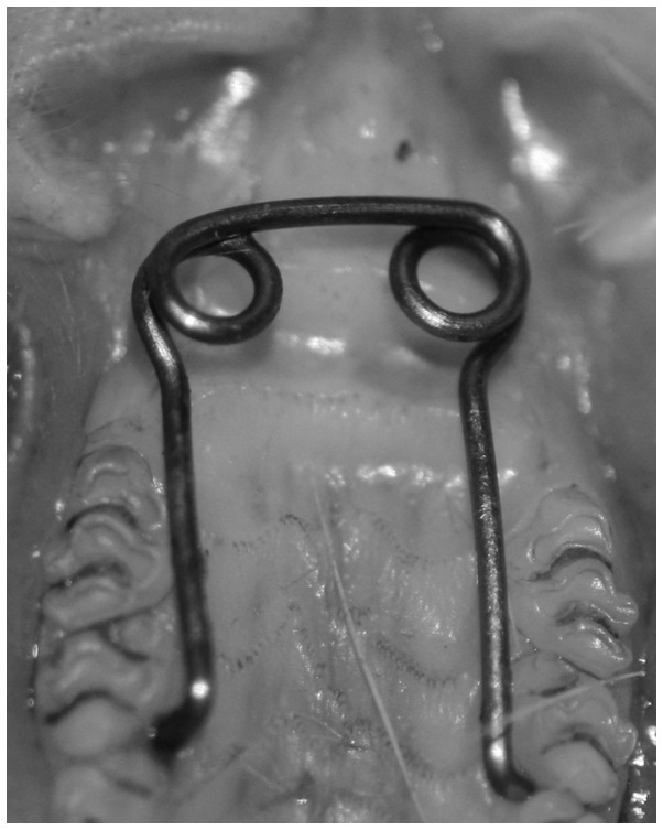
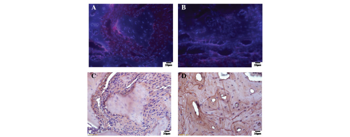
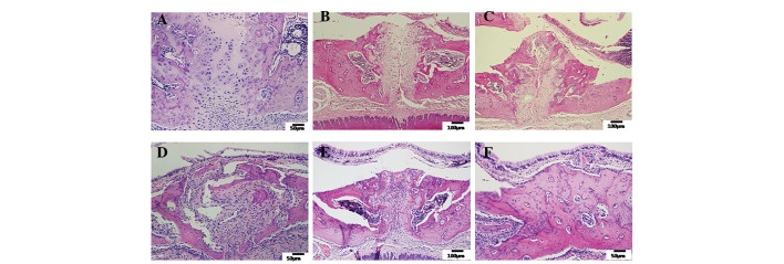
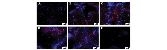
The aim of this study was to explore whether bone marrow mononuclear cell (BMMC) transplantation is able to accelerate the bone remodeling induced by midpalatal expansion in rats. A total of 48 male Sprague-Dawley rats (mean weight, 208.36±7.32 g) were divided into control and midpalatal expansion with or without BMMC transplantation groups. Histological and morphological changes were observed in each group. The osteogenic activities and differential potentials of the transplanted BMMCs labeled with bromodeoxyuridine in the midpalatal bone tissue were assessed by osteocalcin expression. The receptor activator of nuclear factor κB ligand (RANKL)/osteoprotegerin (OPG) ratio was determined by reverse transcription-quantitative polymerase chain reaction (RT-qPCR) to reflect the equilibrium between bone resorption and formation. The results demonstrated that the width of the maxillary dental arch increased distinctly within 2 weeks of midpalatal expansion with BMMC transplantation. The morphology of the midpalatal suture in this group changed significantly; the cartilage was completely replaced by fibrous-like tissue expressing osteocalcin. The palatal bone was reorganized from a cancellous form into a mature compact structure after an additional 2-week relapse period. Immunostaining results indicated that the heterologous transplanted BMMCs survived and differentiated into osteoblasts during the remodeling induced by midpalatal expansion. The RANKL/OPG expression ratio significantly decreased after 2 weeks of midpalatal expansion with BMMC transplantation due to the inhibition of RANKL expression. Heterologous BMMC transplantation appears to accelerate the midpalatal bone remodeling induced by expansion of the rats through increasing the number of osteoprogenitor cells and regulating the RANKL-OPG signaling pathway.
本研究的目的是探讨骨髓单个核细胞(BMMC)移植是否能够加速大鼠腭中缝扩展诱导的骨重塑。总共48只雄性Sprague-Dawley大鼠(平均体重,208.36±7.32 g)被分为对照组以及腭中缝扩展伴或不伴BMMC移植组。观察每组的组织学和形态学变化。通过骨钙素表达评估腭中缝骨组织中用溴脱氧尿苷标记的移植BMMC的成骨活性和分化潜能。通过逆转录定量聚合酶链反应(RT-qPCR)测定核因子κB受体活化因子配体(RANKL)/骨保护素(OPG)比值,以反映骨吸收与形成之间的平衡。结果表明,在进行BMMC移植的腭中缝扩展2周内,上颌牙弓宽度明显增加。该组腭中缝的形态发生了显著变化;软骨完全被表达骨钙素的纤维样组织取代。经过另外2周的复发期后,腭骨从松质骨形式重新组织成成熟的致密结构。免疫染色结果表明,在腭中缝扩展诱导的重塑过程中,异种移植的BMMC存活并分化为成骨细胞。由于RANKL表达受到抑制,在进行BMMC移植的腭中缝扩展2周后,RANKL/OPG表达比值显著降低。异种BMMC移植似乎通过增加骨祖细胞数量和调节RANKL-OPG信号通路来加速大鼠腭中缝扩展诱导的骨重塑。