Liang Ming, Wang Yun, Liang Anlin, Mitch William E, Roy-Chaudhury Prabir, Han Guofeng, Cheng Jizhong
Department of Nephrology, Guangzhou First People's Hospital, Guangzhou Medical University, China.
Division of Nephrology, Department of Medicine, Baylor College of Medicine, Houston, Texas, USA.
Kidney Int. 2015 Sep;88(3):490-502. doi: 10.1038/ki.2015.73. Epub 2015 Mar 18.
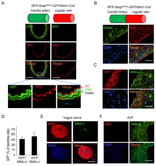
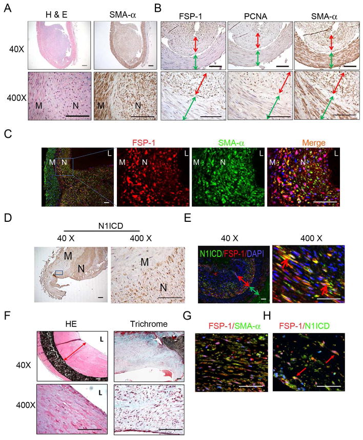
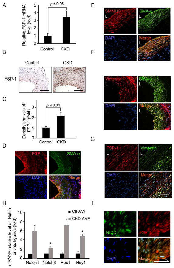
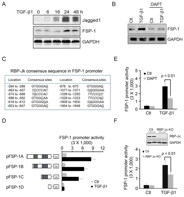
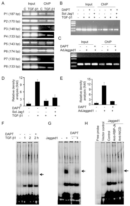
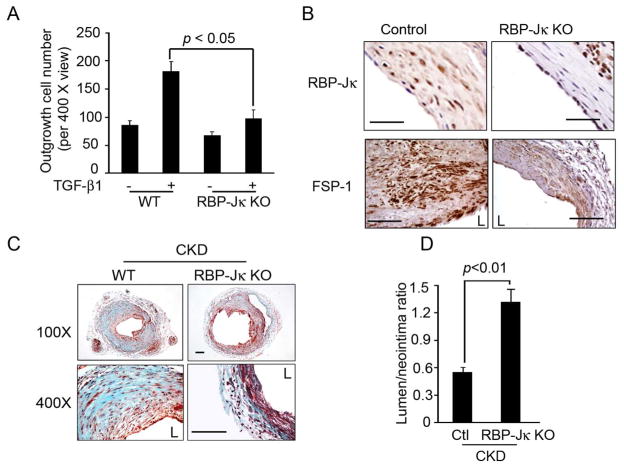
A major factor contributing to failure of arteriovenous fistulas (AVFs) is migration of smooth muscle cells into the forming neointima. To identify the source of smooth muscle cells in neointima, we created end-to-end AVFs by anastomosing the common carotid artery to the jugular vein and studied neural crest-derived smooth muscle cells from the carotid artery, which are Wnt1-positive during development. In Wnt1-cre-GFP mice, smooth muscle cells in the carotid artery but not the jugular vein are labeled with GFP. About half of the cells were GFP-positive in the neointima, indicating their migration from the carotid artery to the jugular vein in AVFs created in these mice. As fibroblast-specific protein-1 (FSP-1) regulates smooth muscle cell migration, we examined FSP-1 in failed AVFs and polytetrafluoroethylene grafts from patients with end-stage kidney disease or from AVFs in mice with chronic kidney disease. In smooth muscle cells of AVFs or polytetrafluoroethylene grafts, FSP-1 and activation of Notch1 are present. In smooth muscle cells, Notch1 increased RBP-Jκ transcription factor activity and RBP-Jκ stimulated FSP-1 expression. Conditional knockout of RBP-Jκ in smooth muscle cells or general knockout of FSP-1 suppressed neointima formation in AVFs in mice. Thus, the artery of AVFs is the major source of smooth muscle cells during neointima formation. Knockout of RBP-Jκ or FSP-1 ameliorates neointima formation and might improve AVF patency during long-term follow-up.
动静脉内瘘(AVF)失败的一个主要因素是平滑肌细胞迁移至正在形成的新生内膜中。为了确定新生内膜中平滑肌细胞的来源,我们通过将颈总动脉与颈静脉吻合创建了端对端AVF,并研究了来自颈动脉的神经嵴衍生平滑肌细胞,这些细胞在发育过程中Wnt1呈阳性。在Wnt1-cre-GFP小鼠中,颈动脉而非颈静脉中的平滑肌细胞被GFP标记。新生内膜中约一半的细胞为GFP阳性,表明它们在这些小鼠创建的AVF中从颈动脉迁移至颈静脉。由于成纤维细胞特异性蛋白-1(FSP-1)调节平滑肌细胞迁移,我们检测了终末期肾病患者的失败AVF和聚四氟乙烯移植物或慢性肾病小鼠AVF中的FSP-1。在AVF或聚四氟乙烯移植物的平滑肌细胞中,存在FSP-1和Notch1的激活。在平滑肌细胞中,Notch1增加了RBP-Jκ转录因子活性,且RBP-Jκ刺激了FSP-1表达。平滑肌细胞中RBP-Jκ的条件性敲除或FSP-1的全身性敲除抑制了小鼠AVF中新生内膜的形成。因此,AVF的动脉是新生内膜形成过程中平滑肌细胞的主要来源。RBP-Jκ或FSP-1的敲除改善了新生内膜形成,并可能在长期随访中提高AVF的通畅率。