Yang Binxia, Janardhanan Rajiv, Vohra Pawan, Greene Eddie L, Bhattacharya Santanu, Withers Sarah, Roy Bhaskar, Nieves Torres Evelyn C, Mandrekar Jaywant, Leof Edward B, Mukhopadhyay Debabrata, Misra Sanjay
Vascular and Interventional Radiology Translational Laboratory, Department of Radiology, Mayo Clinic, Rochester, Minnesota, USA.
Division of Nephrology and Hypertension, Mayo Clinic, Rochester, Minnesota, USA.
Kidney Int. 2014 Feb;85(2):289-306. doi: 10.1038/ki.2013.290. Epub 2013 Aug 7.
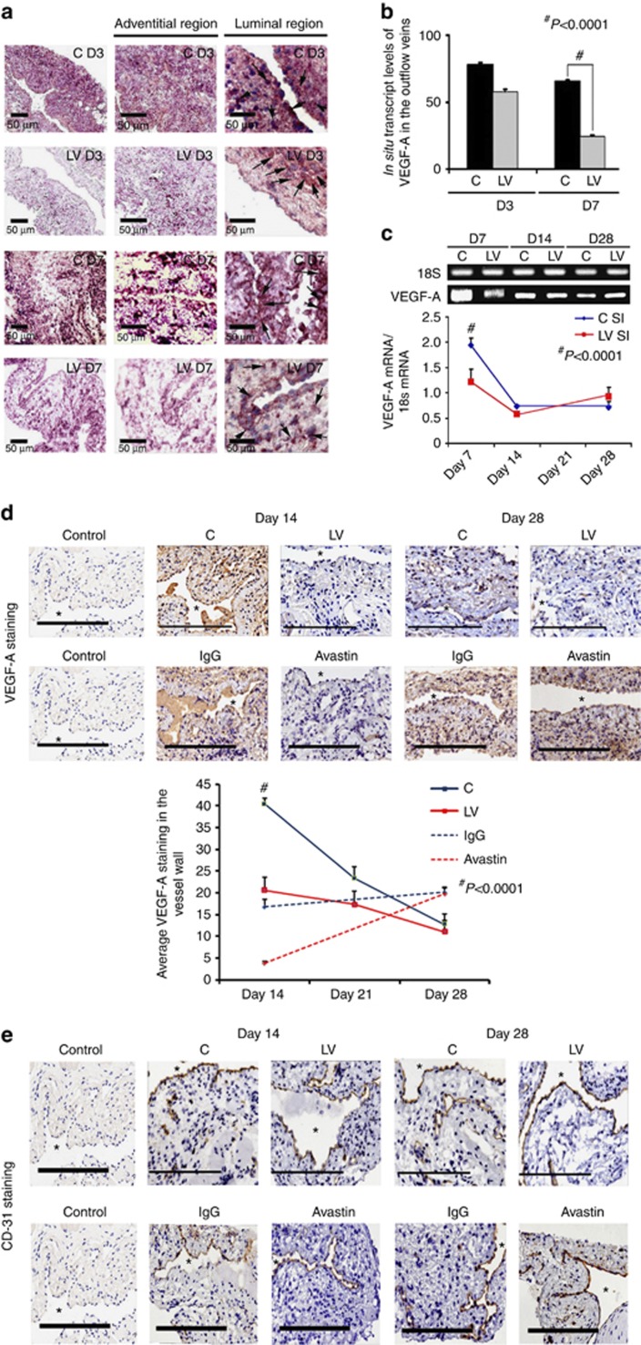
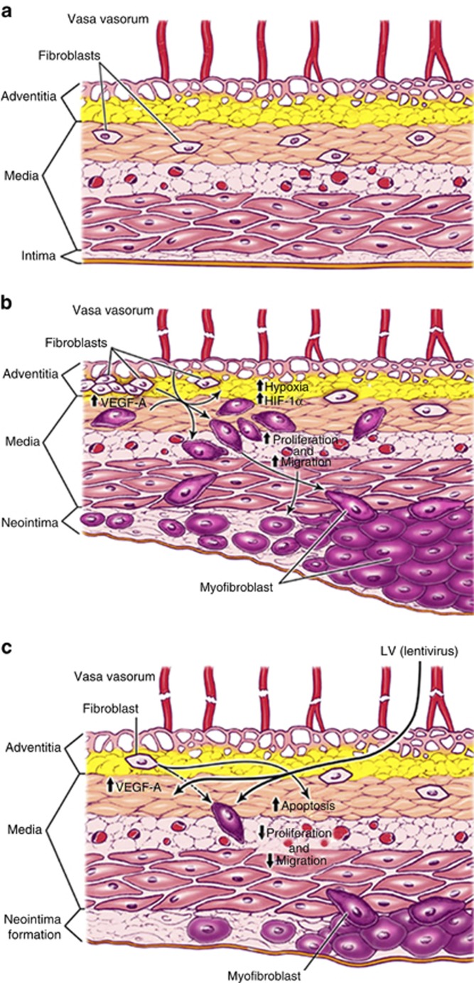
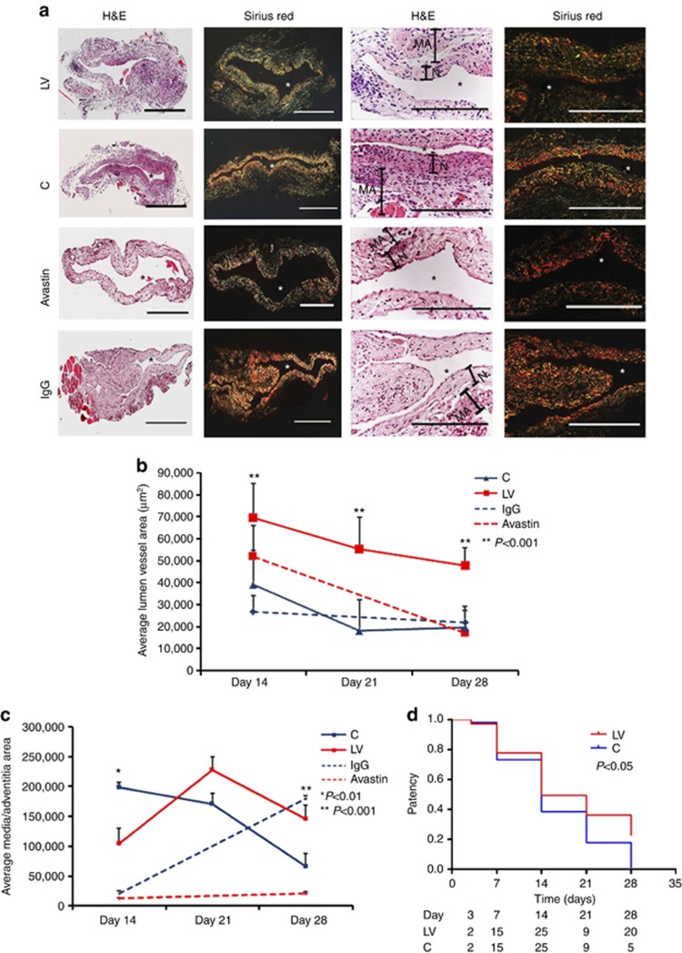
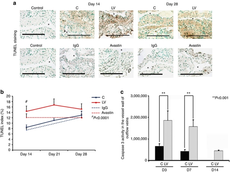
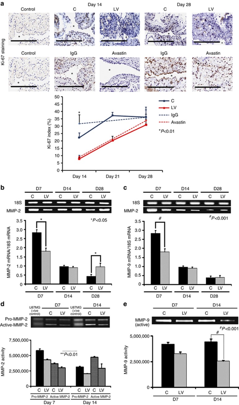
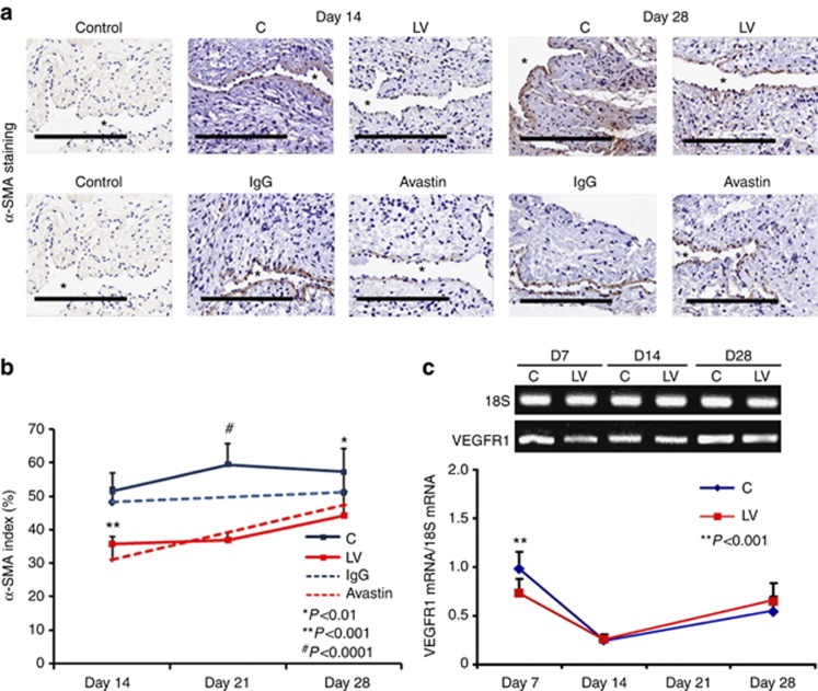
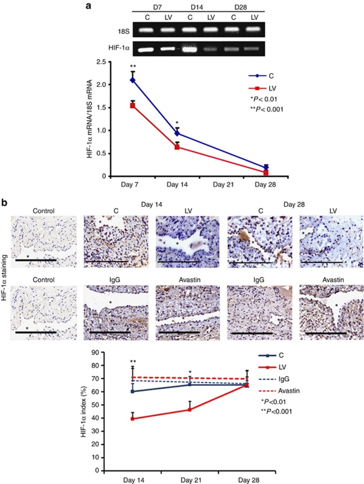
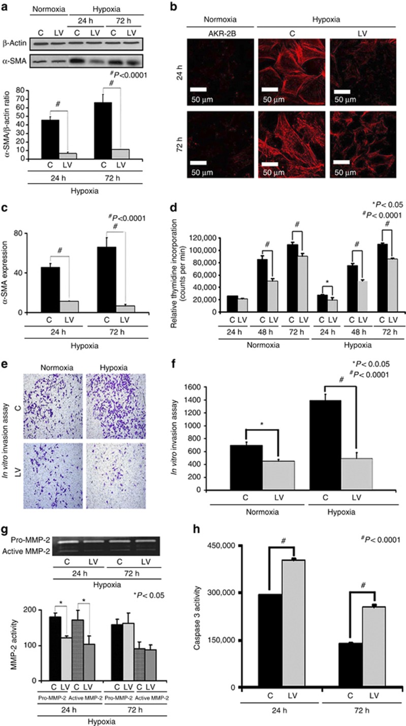
Venous neointimal hyperplasia (VNH) causes hemodialysis vascular access failure. Here we tested whether VNH formation occurs in part due to local vessel hypoxia caused by surgical trauma to the vasa vasorum of the outflow vein at the time of arteriovenous fistula placement. Selective targeting of the adventitia of the outflow vein at the time of fistula creation was performed using a lentivirus-delivered small-hairpin RNA that inhibits VEGF-A expression. This resulted in significant increase in mean lumen vessel area, decreased media/adventitia area, and decreased constrictive remodeling with a significant increase in apoptosis (increase in caspase 3 activity and TUNEL staining) accompanied with decreased cellular proliferation and hypoxia-inducible factor-1α at the outflow vein. There was significant decrease in cells staining positive for α-smooth muscle actin (a myofibroblast marker) and VEGFR-1 expression with a decrease in MMP-2 and MMP-9. These results were confirmed in animals that were treated with humanized monoclonal antibody to VEGF-A with similar results. Since hypoxia can cause fibroblast to differentiate into myofibroblasts, we silenced VEGF-A gene expression in fibroblasts and subjected them to hypoxia. This decreased myofibroblast production, cellular proliferation, cell invasion, MMP-2 activity, and increased caspase 3. Thus, VEGF-A reduction at the time of arteriovenous fistula placement results in increased positive vascular remodeling.
静脉内膜增生(VNH)会导致血液透析血管通路失败。在此,我们测试了VNH的形成是否部分归因于动静脉内瘘置入时,流出静脉的血管滋养管受到手术创伤所导致的局部血管缺氧。在造瘘时,使用携带抑制VEGF-A表达的小发夹RNA的慢病毒,对流出静脉的外膜进行选择性靶向。这导致平均管腔面积显著增加、中膜/外膜面积减小、收缩性重塑减少,同时凋亡显著增加(半胱天冬酶3活性和TUNEL染色增加),伴有流出静脉处细胞增殖和缺氧诱导因子-1α减少。α-平滑肌肌动蛋白(一种肌成纤维细胞标志物)染色阳性的细胞和VEGFR-1表达显著减少,同时MMP-2和MMP-9也减少。在用抗VEGF-A人源化单克隆抗体治疗的动物中也证实了这些结果,且结果相似。由于缺氧可导致成纤维细胞分化为肌成纤维细胞,我们使成纤维细胞中的VEGF-A基因表达沉默,并使其处于缺氧状态。这减少了肌成纤维细胞的产生、细胞增殖、细胞侵袭、MMP-2活性,并增加了半胱天冬酶3。因此,动静脉内瘘置入时VEGF-A的减少会导致血管正向重塑增加。