Utzinger Urs, Baggett Brenda, Weiss Jeffrey A, Hoying James B, Edgar Lowell T
Department of Biomedical Engineering, University of Arizona, Tucson, AZ, USA,
Angiogenesis. 2015 Jul;18(3):219-32. doi: 10.1007/s10456-015-9461-x. Epub 2015 Mar 21.
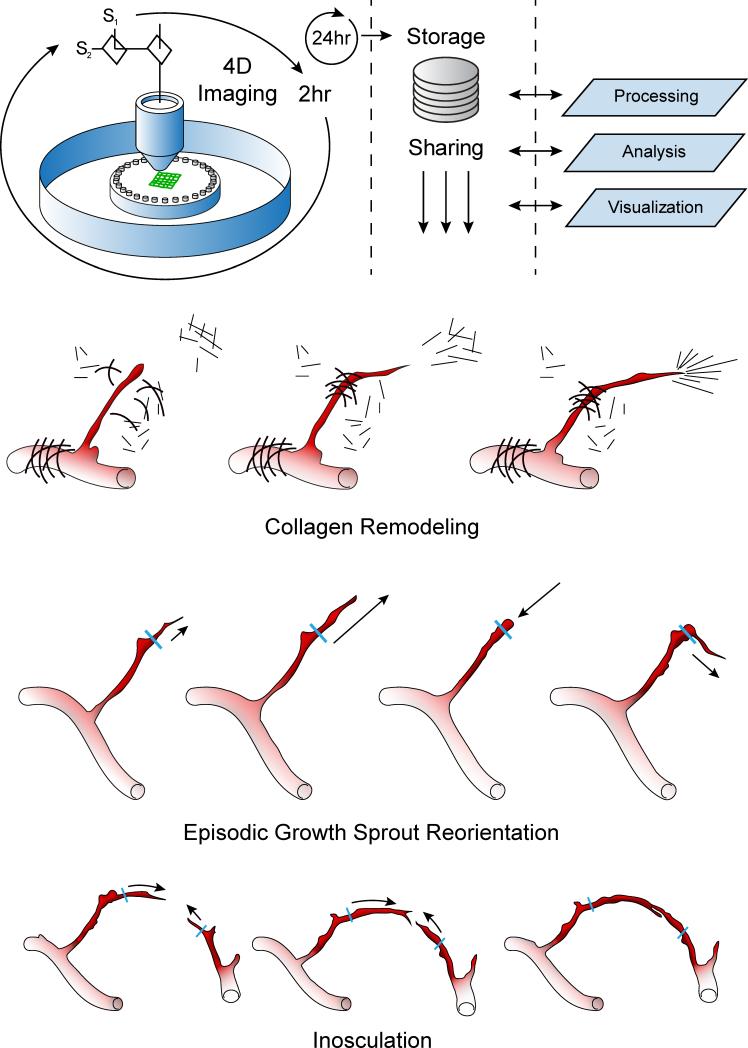
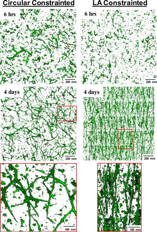
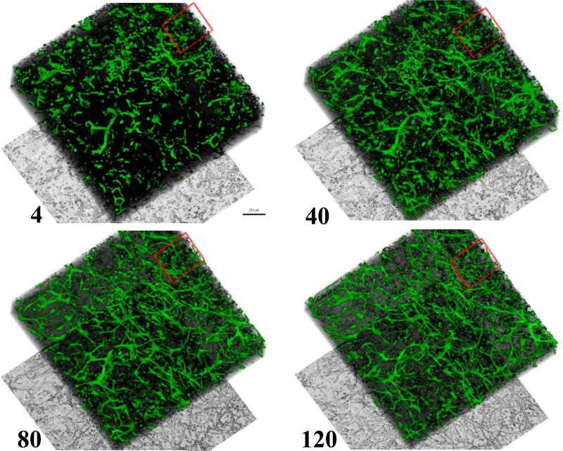
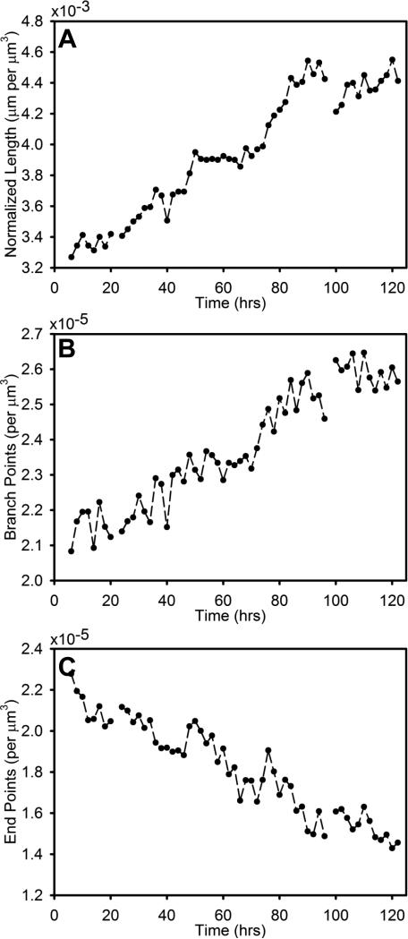
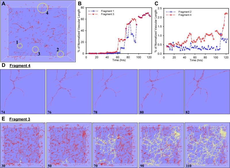
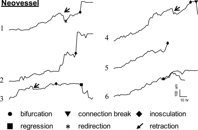
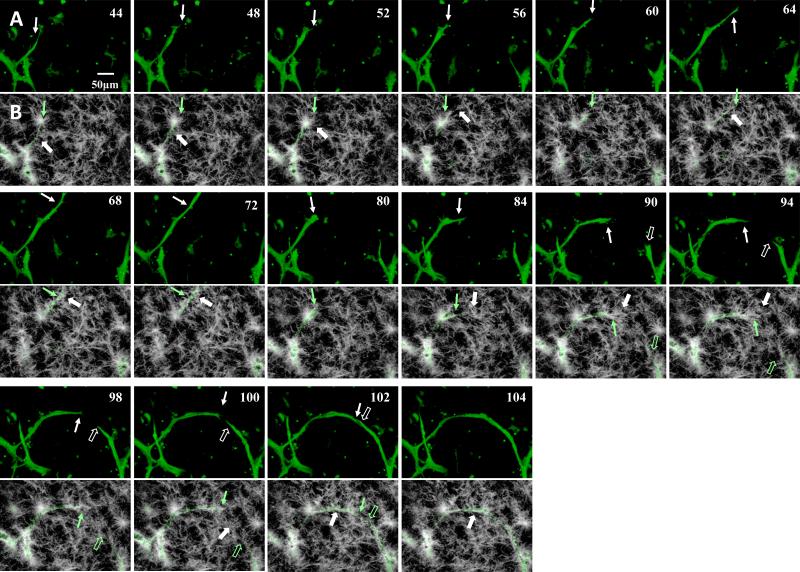
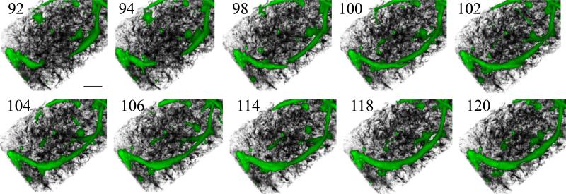
During angiogenesis, growing neovessels must effectively navigate through the tissue space as they elongate and subsequently integrate into a microvascular network. While time series microscopy has provided insight into the cell activities within single growing neovessel sprouts, less is known concerning neovascular dynamics within a large angiogenic tissue bed. Here, we developed a time-lapse imaging technique that allowed visualization and quantification of sprouting neovessels as they form and grow away from adult parent microvessels in three dimensions over cubic millimeters of matrix volume during the course of up to 5 days on the microscope. Using a new image acquisition procedure and novel morphometric analysis tools, we quantified the elongation dynamics of growing neovessels and found an episodic growth pattern accompanied by fluctuations in neovessel diameter. Average elongation rate was 5 μm/h for individual vessels, but we also observed considerable dynamic variability in growth character including retraction and complete regression of entire neovessels. We observed neovessel-to-neovessel directed growth over tens to hundreds of microns preceding tip-to-tip inosculation. As we have previously described via static 3D imaging at discrete time points, we identified different collagen fibril structures associated with the growing neovessel tip and stalk, and observed the coordinated alignment of growing neovessels in a deforming matrix. Overall analysis of the entire image volumes demonstrated that although individual neovessels exhibited episodic growth and regression, there was a monotonic increase in parameters associated with the entire vascular bed such as total network length and number of branch points. This new time-lapse imaging approach corroborated morphometric changes in individual neovessels described by us and others, as well as captured dynamic neovessel behaviors unique to days-long angiogenesis within the forming neovascular network.
在血管生成过程中,新生血管在伸长并随后整合到微血管网络的过程中,必须有效地在组织空间中穿行。虽然时间序列显微镜已经让我们深入了解了单个生长中的新生血管芽内的细胞活动,但对于大型血管生成组织床内的新生血管动力学却知之甚少。在此,我们开发了一种延时成像技术,该技术能够在显微镜下对新生血管从成年亲本微血管形成并在三维空间中于立方毫米的基质体积内生长长达5天的过程进行可视化和定量分析。通过使用一种新的图像采集程序和新颖的形态计量分析工具,我们对生长中的新生血管的伸长动力学进行了量化,发现了一种伴有新生血管直径波动的间歇性生长模式。单个血管的平均伸长率为5μm/h,但我们也观察到生长特征存在相当大的动态变异性,包括整个新生血管的回缩和完全消退。在尖端对尖端吻合之前,我们观察到新生血管之间在数十到数百微米范围内的定向生长。正如我们之前在离散时间点通过静态三维成像所描述的那样,我们识别出了与生长中的新生血管尖端和茎干相关的不同胶原纤维结构,并观察到生长中的新生血管在变形基质中的协同排列。对整个图像体积的总体分析表明,尽管单个新生血管表现出间歇性生长和消退,但与整个血管床相关的参数,如总网络长度和分支点数,却呈单调增加。这种新的延时成像方法证实了我们和其他人所描述的单个新生血管的形态计量变化,同时也捕捉到了在形成的新生血管网络中长达数天的血管生成过程中独特的新生血管动态行为。