de Oliveira Patricia Bedesco, Puchnick Andrea, Szejnfeld Jacob, Goldman Suzan Menasce
Department of Imaging Diagnosis, Escola Paulista de Medicina, Universidade Federal de São Paulo (UNIFESP), São Paulo, SP, Brazil.
PLoS One. 2015 Mar 23;10(3):e0121317. doi: 10.1371/journal.pone.0121317. eCollection 2015.
To ascertain the prevalence of pancreatic cysts detected incidentally on 3-Tesla magnetic resonance imaging (MRI) of the abdomen and correlate this prevalence with patient age and gender; assess the number, location, and size of these lesions, as well as features suspicious for malignancy; and determine the prevalence of incidentally detected dilatation of the main pancreatic duct (MPD).
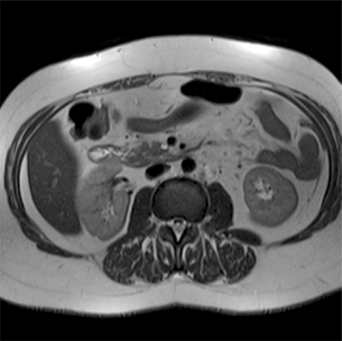
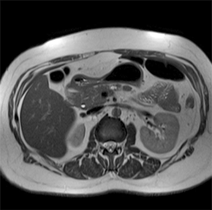
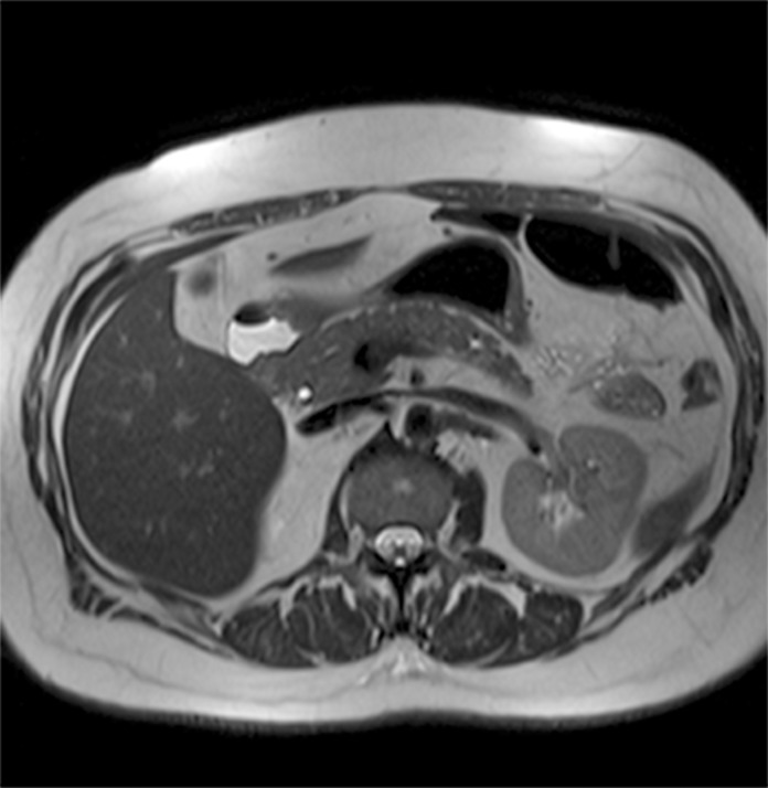
Retrospective analysis of 2,678 reports of patients who underwent abdominal MRI between January 2012 and June 2013. Patients with a known history of pancreatic conditions or surgery were excluded, and the remaining 2,583 reports were examined for the presence of pancreatic cysts, which was then correlated with patient age and gender. We also assessed whether cysts were solitary or multiple, as well as their location within the pancreatic parenchyma, size, and features suspicious for malignancy. Finally, we calculated the prevalence of incidental MPD dilatation, defined as MPD diameter ≥ 2.5 mm.
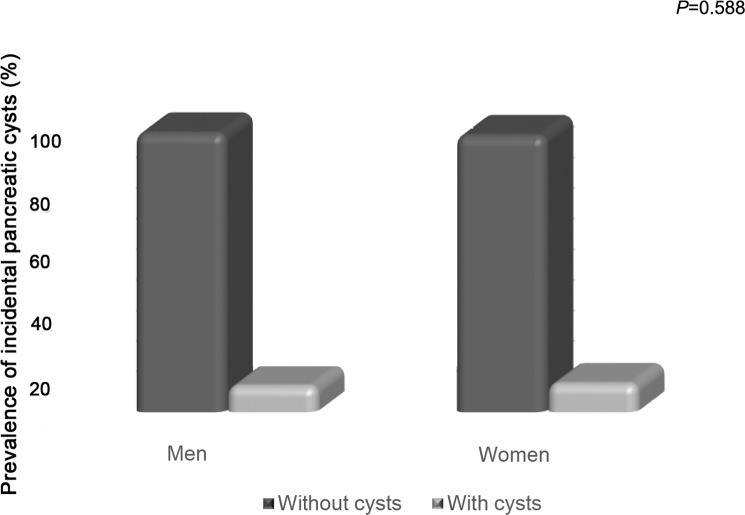
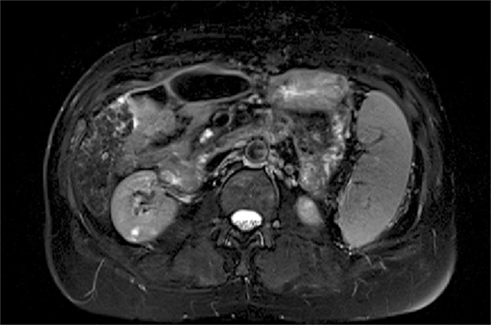
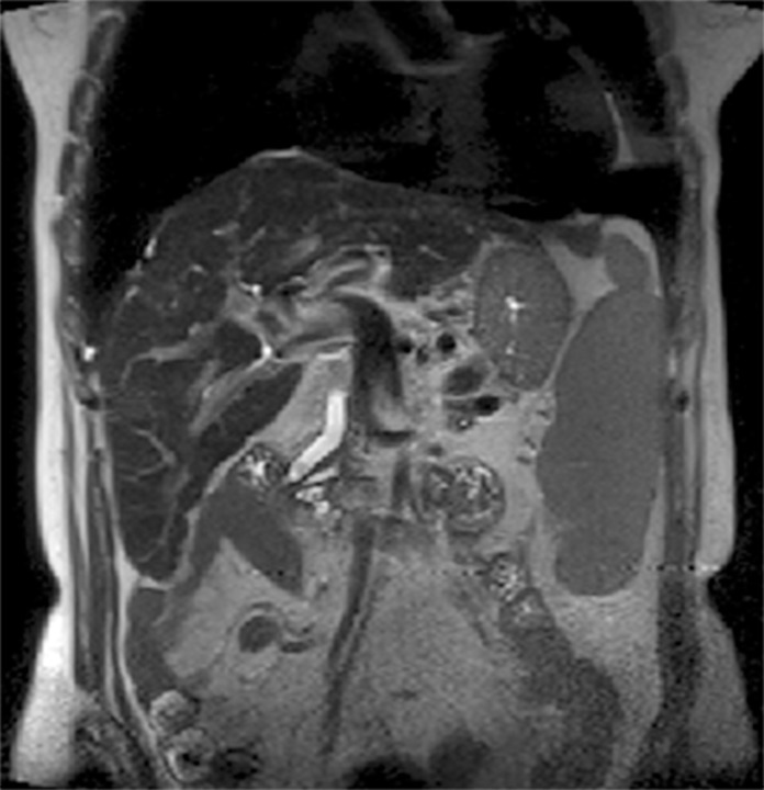
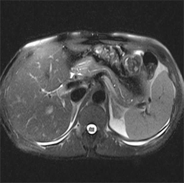
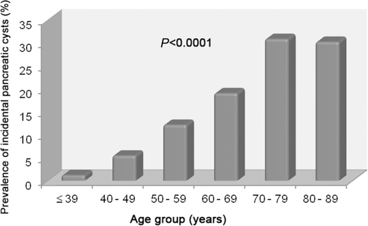
Pancreatic cysts were detected incidentally in 9.3% of patients (239/2,583). The prevalence of pancreatic cysts increased significantly with age (p<0.0001). There were no significant differences in prevalence between men and women (p=0.588). Most cysts were multiple (57.3%), distributed diffusely throughout the pancreas (41.8%), and 5 mm or larger (81.6%). In 12.1% of cases, cysts exhibited features suspicious for malignancy. Overall, 2.7% of subjects exhibited incidental MPD dilatation.
In this sample, the prevalence of pancreatic cysts detected incidentally on 3T MRI of the abdomen was 9.3%. Prevalence increased with age and was not associated with gender. The majority of cysts were multiple, diffusely distributed through the pancreatic parenchyma, and ≥ 5 mm in size; 12.1% were suspicious for malignancy. An estimated 2.7% of subjects had a dilated MPD.
确定在腹部3特斯拉磁共振成像(MRI)检查中偶然发现的胰腺囊肿的患病率,并将该患病率与患者年龄和性别相关联;评估这些病变的数量、位置和大小,以及可疑恶性特征;并确定偶然发现的主胰管(MPD)扩张的患病率。
回顾性分析2012年1月至2013年6月期间接受腹部MRI检查的2678例患者的报告。排除有已知胰腺疾病或手术史的患者,对其余2583份报告检查是否存在胰腺囊肿,然后将其与患者年龄和性别相关联。我们还评估囊肿是单发还是多发,以及它们在胰腺实质内的位置、大小和可疑恶性特征。最后,我们计算偶然MPD扩张的患病率,定义为MPD直径≥2.5毫米。
9.3%的患者(239/2583)偶然发现胰腺囊肿。胰腺囊肿的患病率随年龄显著增加(p<0.0001)。男性和女性的患病率无显著差异(p=0.588)。大多数囊肿为多发(57.3%),弥漫分布于整个胰腺(41.8%),且直径为5毫米或更大(81.6%)。在12.1%的病例中,囊肿表现出可疑恶性特征。总体而言,2.7%的受试者表现出偶然的MPD扩张。
在本样本中,腹部3T MRI偶然发现的胰腺囊肿患病率为9.3%。患病率随年龄增加,与性别无关。大多数囊肿为多发,弥漫分布于胰腺实质,大小≥5毫米;12.1%可疑恶性。估计2.7%的受试者有MPD扩张。