Carter Teralyn E, Taylor Kevin A, Spritzer Charles E, Utturkar Gangadhar M, Taylor Dean C, Moorman Claude T, Garrett William E, Guilak Farshid, McNulty Amy L, DeFrate Louis E
Department of Orthopaedic Surgery, Duke University Medical Center, Durham, NC, United States.
Department of Radiology, Duke University Medical Center, Durham, NC, United States.
J Biomech. 2015 Jun 1;48(8):1461-8. doi: 10.1016/j.jbiomech.2015.02.030. Epub 2015 Mar 5.
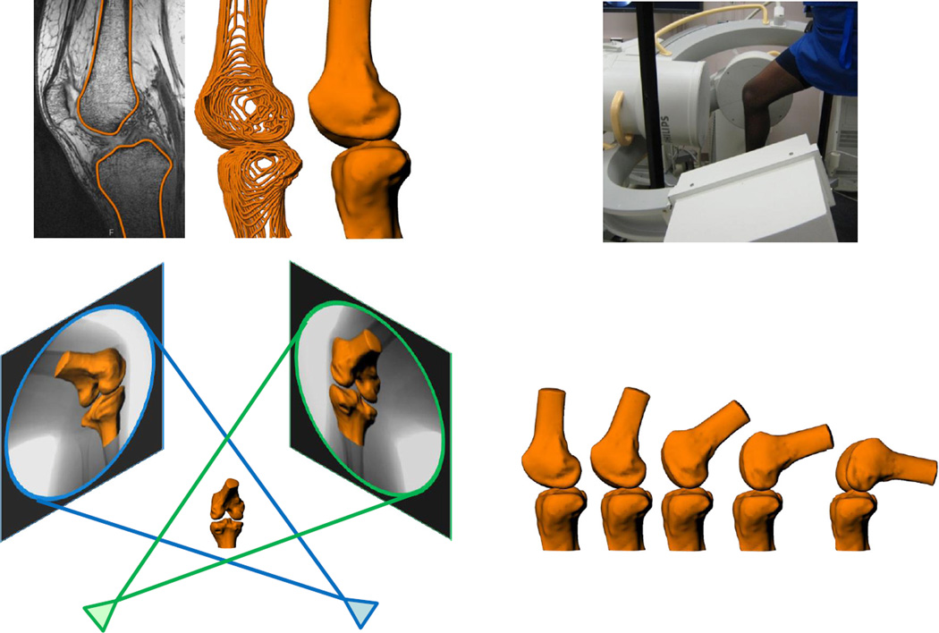
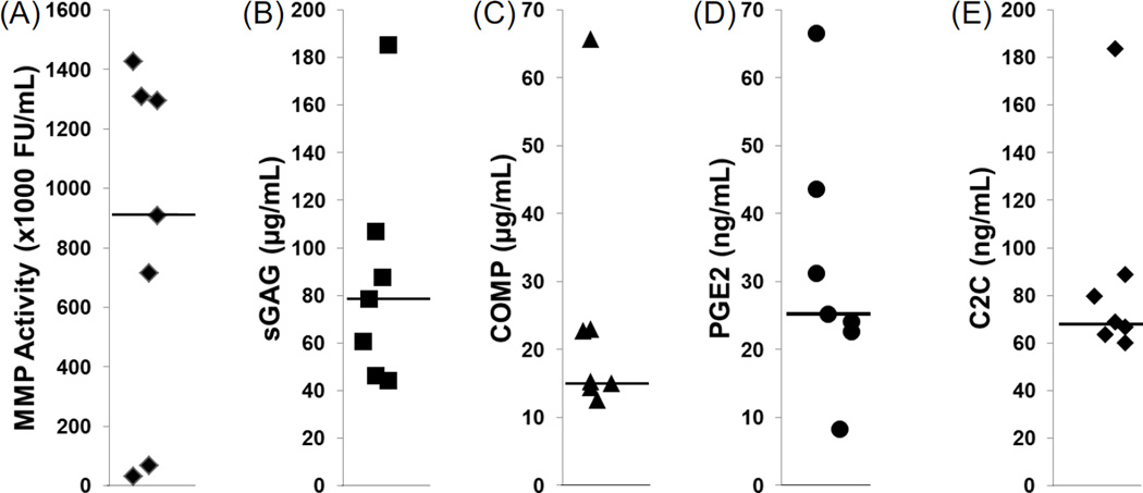
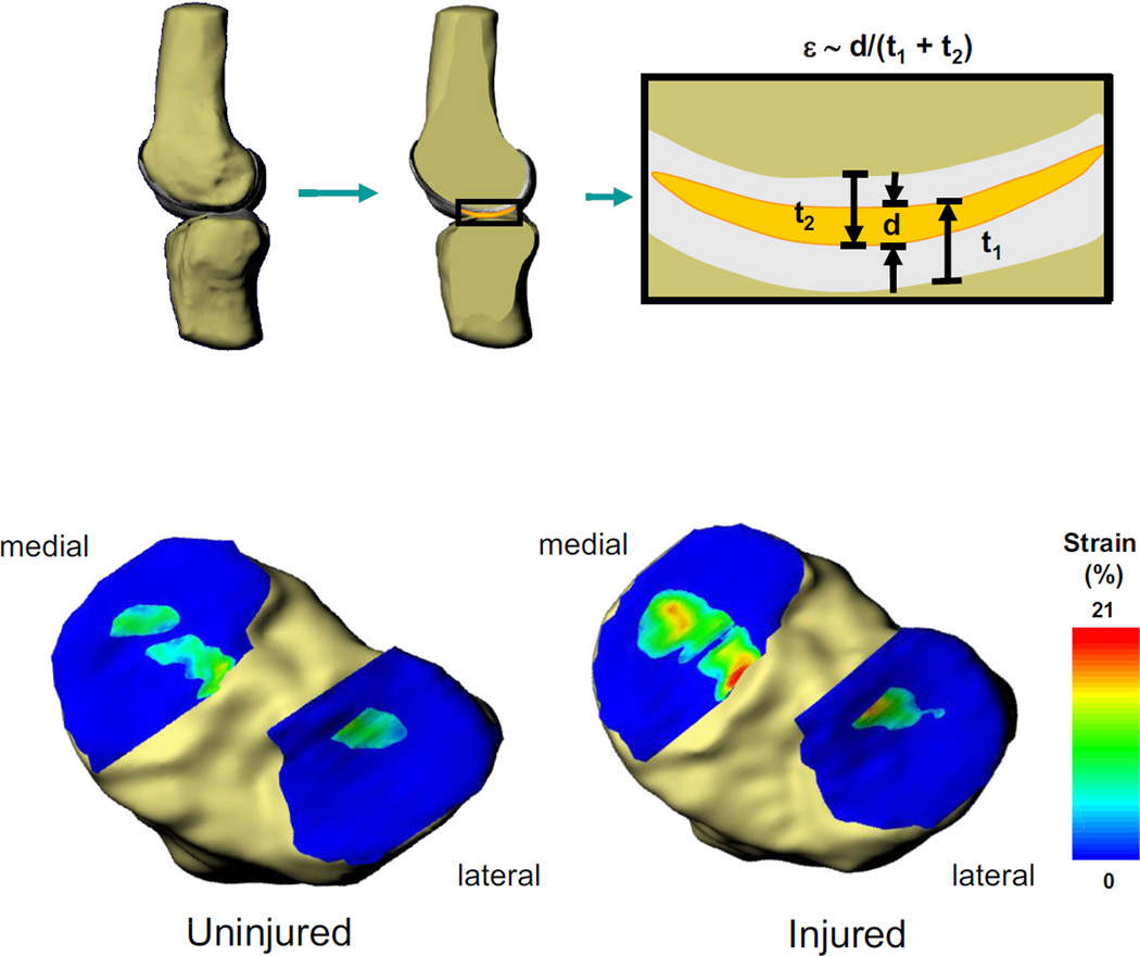
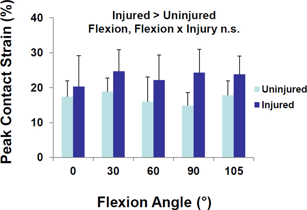
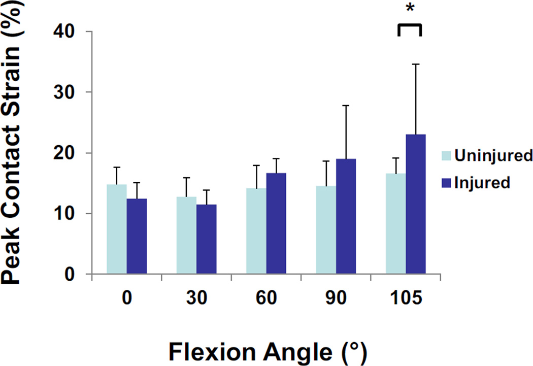
Meniscal tears are common injuries, and while partial meniscectomy is a frequent treatment option, general meniscus loss is a risk factor for the development of osteoarthritis. The goal of this study was to measure the in vivo tibiofemoral cartilage contact patterns in patients with meniscus tears in relation to biomarkers of cartilage catabolism in the synovial fluid of these joints. A combination of magnetic resonance imaging and biplanar fluoroscopy was used to determine the in vivo motion and cartilage contact mechanics of the knee. Subjects with isolated medial meniscus tears were analyzed while performing a quasi-static lunge, and the contralateral uninjured knee was used as a control. Synovial fluid was collected from the injured knee and matrix metalloproteinase (MMP) activity, sulfated glycosaminoglycan, cartilage oligomeric matrix protein, prostaglandin E2, and the collagen type II cleavage biomarker C2C were measured. Contact strain in the medial compartment increased significantly in the injured knees compared to contralateral control knees. In the lateral compartment, the contact strain in the injured knee was significantly increased only at the maximum flexion angle (105°). The average cartilage strain at maximum flexion positively correlated with total MMP activity in the synovial fluid. These findings show that meniscal injury leads to loss of normal joint function and increased strain of the articular cartilage, which correlated to elevated total MMP activity in the synovial fluid. The increased strain and total MMP activity may reflect, or potentially contribute to, the early development of osteoarthritis that is observed following meniscal injury.
半月板撕裂是常见的损伤,虽然部分半月板切除术是常用的治疗选择,但半月板整体缺失是骨关节炎发生的一个风险因素。本研究的目的是测量半月板撕裂患者体内胫股关节软骨的接触模式,并将其与这些关节滑液中软骨分解代谢的生物标志物相关联。结合磁共振成像和双平面荧光透视法来确定膝关节的体内运动和软骨接触力学。对孤立性内侧半月板撕裂的受试者在进行准静态弓步动作时进行分析,对侧未受伤的膝关节作为对照。从受伤膝关节收集滑液,测量基质金属蛋白酶(MMP)活性、硫酸化糖胺聚糖、软骨寡聚基质蛋白、前列腺素E2以及II型胶原裂解生物标志物C2C。与对侧对照膝关节相比,受伤膝关节内侧间室的接触应变显著增加。在外侧间室,受伤膝关节的接触应变仅在最大屈曲角度(105°)时显著增加。最大屈曲时的平均软骨应变与滑液中MMP总活性呈正相关。这些发现表明,半月板损伤导致正常关节功能丧失和关节软骨应变增加,这与滑液中MMP总活性升高相关。应变增加和MMP总活性升高可能反映或潜在地促成了半月板损伤后观察到的骨关节炎的早期发展。