Baldini Enke, Tuccilli Chiara, Prinzi Natalie, Sorrenti Salvatore, Falvo Laura, De Vito Corrado, Catania Antonio, Tartaglia Francesco, Mocini Renzo, Coccaro Carmela, Alessandrini Stefania, Barollo Susi, Mian Caterina, Antonelli Alessandro, De Antoni Enrico, D'Armiento Massimino, Ulisse Salvatore
Department of Experimental Medicine, "Sapienza" University of Rome, Rome, Italy.
Department of Surgical Sciences, "Sapienza" University of Rome, Rome, Italy.
PLoS One. 2015 Mar 25;10(3):e0121514. doi: 10.1371/journal.pone.0121514. eCollection 2015.
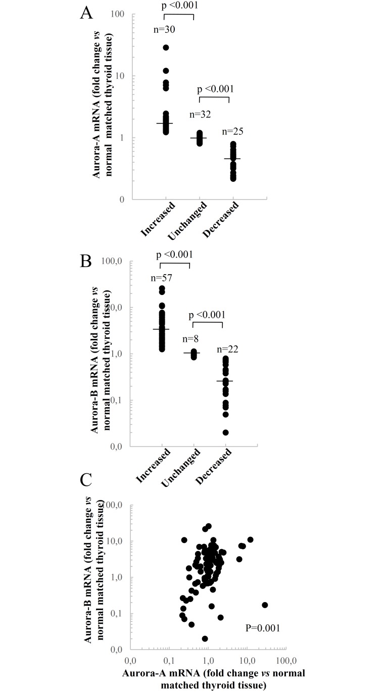
A number of reports indicated that Aurora-A or Aurora-B overexpression represented a negative prognostic factor in several human malignancies. In thyroid cancer tissues a deregulated expression of Aurora kinases has been also demonstrated, but no information regarding its possible prognostic role in differentiated thyroid cancer is available. Here, we evaluated Aurora-A and Aurora-B mRNA expression and its prognostic relevance in a series of 87 papillary thyroid cancers (PTC), with a median follow-up of 63 months. The analysis of Aurora-A and Aurora-B mRNA levels in PTC tissues, compared to normal matched tissues, revealed that their expression was either up- or down-regulated in the majority of cancer tissues. In particular, Aurora-A and Aurora-B mRNA levels were altered, respectively, in 55 (63.2%) and 79 (90.8%) out of the 87 PTC analyzed.A significant positive correlation between Aurora-A and Aurora-B mRNAs was observed (p=0.001). The expression of both Aurora genes was not affected by the BRAFV600E mutation. Univariate, multivariate and Kaplan-Mayer analyses documented the lack of association between Aurora-A or Aurora-B expression and clinicopathological parameters such as gender, age, tumor size, histology, TNM stage, lymph node metastasis and BRAF status as well as disease recurrences or disease-free interval. Only Aurora-B mRNA was significantly higher in T(3-4) tissues, with respect to T(1-2) PTC tissues. The data reported here demonstrate that the expression of Aurora kinases is deregulated in the majority of PTC tissues, likely contributing to PTC progression. However, differently from other human solid cancers, detection of Aurora-A or Aurora-B mRNAs is not a prognostic biomarker in PTC patients.
多项报告表明,极光激酶A(Aurora-A)或极光激酶B(Aurora-B)的过表达在多种人类恶性肿瘤中是一个负面预后因素。在甲状腺癌组织中也已证实极光激酶表达失调,但关于其在分化型甲状腺癌中可能的预后作用尚无相关信息。在此,我们评估了87例乳头状甲状腺癌(PTC)中极光激酶A和极光激酶B的mRNA表达及其预后相关性,中位随访时间为63个月。与正常匹配组织相比,对PTC组织中极光激酶A和极光激酶B的mRNA水平分析显示,它们在大多数癌组织中的表达要么上调要么下调。特别是,在分析的87例PTC中,分别有55例(63.2%)和79例(90.8%)的极光激酶A和极光激酶B的mRNA水平发生改变。观察到极光激酶A和极光激酶B的mRNA之间存在显著正相关(p = 0.001)。两种极光基因的表达均不受BRAFV600E突变的影响。单因素、多因素和Kaplan-Meier分析表明,极光激酶A或极光激酶B的表达与临床病理参数如性别、年龄、肿瘤大小、组织学、TNM分期、淋巴结转移和BRAF状态以及疾病复发或无病间期之间缺乏关联。仅在T(3 - 4)期组织中,相对于T(1 - 2)期PTC组织,极光激酶B的mRNA显著更高。此处报告的数据表明,极光激酶的表达在大多数PTC组织中失调,可能促进PTC进展。然而,与其他人类实体癌不同,检测极光激酶A或极光激酶B的mRNA并非PTC患者的预后生物标志物。