• 文献检索
  • 文档翻译
  • 深度研究
  • 学术资讯
  • Suppr Zotero 插件Zotero 插件
  • 邀请有礼
  • 套餐&价格
  • 历史记录
应用&插件
Suppr Zotero 插件Zotero 插件浏览器插件Mac 客户端Windows 客户端微信小程序
定价
高级版会员购买积分包购买API积分包
服务
文献检索文档翻译深度研究API 文档MCP 服务
关于我们
关于 Suppr公司介绍联系我们用户协议隐私条款
关注我们

Suppr 超能文献

核心技术专利:CN118964589B侵权必究
粤ICP备2023148730 号-1Suppr @ 2026

文献检索

告别复杂PubMed语法,用中文像聊天一样搜索,搜遍4000万医学文献。AI智能推荐,让科研检索更轻松。

立即免费搜索

文件翻译

保留排版,准确专业,支持PDF/Word/PPT等文件格式,支持 12+语言互译。

免费翻译文档

深度研究

AI帮你快速写综述,25分钟生成高质量综述,智能提取关键信息,辅助科研写作。

立即免费体验

使用T1ρ映射磁共振成像检测与半月板撕裂相关的早期软骨退变

Detection of early cartilage deterioration associated with meniscal tear using T1ρ mapping magnetic resonance imaging.

作者信息

Matsubara Hirokazu, Okazaki Ken, Takayama Yukihisa, Osaki Kanji, Matsuo Yoshio, Honda Hiroshi, Iwamoto Yukihide

出版信息

BMC Musculoskelet Disord. 2015 Feb 10;16(1):22. doi: 10.1186/s12891-015-0487-4.

DOI:10.1186/s12891-015-0487-4
PMID:25808907
原文链接:https://pmc.ncbi.nlm.nih.gov/articles/PMC4327971/
Abstract

BACKGROUND

In patients with degenerative meniscal tears, subclinical cartilage degeneration may be present even if gross morphological changes are not evident. The aim of this study was to detect occult cartilage degeneration using T1ρ MRI mapping in patients with meniscal tears without obvious radiographic osteoarthritis (OA).

METHODS

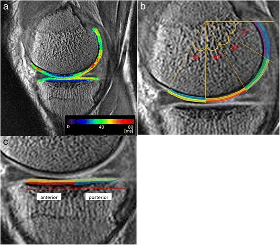
A total of 22 subjects with degenerative meniscal tears in the early stages of osteoarthritis [Kellgren-Lawrence (KL) grade of 0-2] and 19 healthy subjects as the control group were examined. The femoral condyle was divided into four 30° wedges (-30°-0° anteriorly, 0°-30°, 30°-60° and 60°-90° posteriorly), and each area of cartilage was further divided into superficial and deep layers of equal thickness. The tibial side was divided into anterior and posterior areas with superficial and deep layers in each. The mean T1ρ values (ms) in each area were calculated.

RESULTS

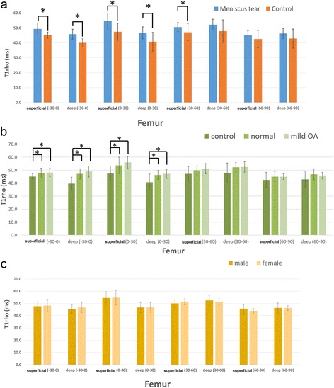
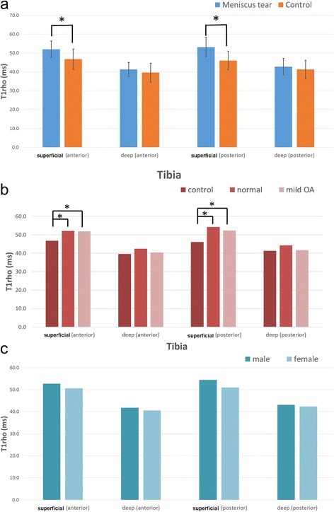
On the femoral side, T1ρ values of the superficial and deep regions (-30°-0°, 0°-30° and 30°-60°) in the meniscal tear group were significantly higher than those in the control group [superficial (-30°-0°): 49.0 ± 4.0 (meniscal tear group) vs 45.1 ± 2.1 (control group), deep (-30°-0°): 45.2 ± 3.3 vs 39.5 ± 5.0, superficial (0°-30°): 54.5 ± 5.3 vs 47.4 ± 5.7, deep (0°-30°): 46.8 ± 4.0 vs 40.7 ± 6.3, superficial (30°-60°): 50.5 ± 3.1 vs 47.1 ± 5.7]. On the tibial side, the meniscal tear group had significantly higher T1ρ values superficially in both anterior and posterior regions compared with the control group [superficial (anterior): 52.0 ± 4.3 vs 46.7 ± 5.4, superficial (posterior): 53.1 ± 5.1 vs 46.0 ± 4.9]. Moreover, these significant differences were observed when comparing patients in the meniscal tear group with KL grades of 0 or 1 and the control group.

CONCLUSIONS

Our study suggested that early biochemical changes in cartilage associated with degenerative meniscal tears occur first in the superficial zones in areas of contact during slight flexion. Characterising the early relationship between cartilage degeneration and degenerative meniscal tears using T1ρ MRI mapping may be of clinical benefit and provide further evidence linking meniscal injury to OA.

摘要

背景

在退行性半月板撕裂患者中,即使没有明显的大体形态学改变,也可能存在亚临床软骨退变。本研究的目的是使用T1ρ磁共振成像(MRI)映射技术检测无明显影像学骨关节炎(OA)的半月板撕裂患者的隐匿性软骨退变。

方法

共检查了22例处于骨关节炎早期(Kellgren-Lawrence(KL)分级为0-2级)的退行性半月板撕裂患者以及19名健康受试者作为对照组。股骨髁被分为四个30°楔形区域(前方-30°至0°,0°至30°,30°至60°以及后方60°至90°),每个软骨区域进一步分为厚度相等的浅层和深层。胫骨侧分为前后区域,每个区域又分为浅层和深层。计算每个区域的平均T1ρ值(毫秒)。

结果

在股骨侧,半月板撕裂组的浅层和深层区域(-30°至0°,0°至30°和30°至60°)的T1ρ值显著高于对照组[浅层(-30°至0°):49.0±4.0(半月板撕裂组)对45.1±2.1(对照组),深层(-30°至0°):45.2±3.3对39.5±5.0,浅层(0°至30°):54.5±5.3对47.4±5.7,深层(0°至30°):46.8±4.0对40.7±6.3,浅层(30°至60°):50.5±3.1对47.1±5.7]。在胫骨侧,半月板撕裂组的前后区域浅层T1ρ值均显著高于对照组[浅层(前方):52.0±4.3对46.7±5.4,浅层(后方):53.1±5.1对46.0±4.9]。此外,在比较KL分级为0或1的半月板撕裂组患者与对照组时也观察到了这些显著差异。

结论

我们的研究表明,与退行性半月板撕裂相关的软骨早期生化变化首先发生在轻微屈曲时接触区域的浅层。使用T1ρ MRI映射技术表征软骨退变与退行性半月板撕裂之间的早期关系可能具有临床益处,并为半月板损伤与OA之间的联系提供进一步证据。

https://cdn.ncbi.nlm.nih.gov/pmc/blobs/a0a3/4327971/d831009a8f5d/12891_2015_487_Fig4_HTML.jpg
https://cdn.ncbi.nlm.nih.gov/pmc/blobs/a0a3/4327971/26a10fdae033/12891_2015_487_Fig1_HTML.jpg
https://cdn.ncbi.nlm.nih.gov/pmc/blobs/a0a3/4327971/11dbda7ea212/12891_2015_487_Fig2_HTML.jpg
https://cdn.ncbi.nlm.nih.gov/pmc/blobs/a0a3/4327971/ab2166bb58c9/12891_2015_487_Fig3_HTML.jpg
https://cdn.ncbi.nlm.nih.gov/pmc/blobs/a0a3/4327971/d831009a8f5d/12891_2015_487_Fig4_HTML.jpg

相似文献

1
Detection of early cartilage deterioration associated with meniscal tear using T1ρ mapping magnetic resonance imaging.使用T1ρ映射磁共振成像检测与半月板撕裂相关的早期软骨退变
BMC Musculoskelet Disord. 2015 Feb 10;16(1):22. doi: 10.1186/s12891-015-0487-4.
2
Articular cartilage lesions increase early cartilage degeneration in knees treated by anterior cruciate ligament reconstruction: T1ρ mapping evaluation and 1-year follow-up.关节软骨损伤增加前交叉韧带重建治疗膝关节的早期软骨退变:T1ρ 图谱评估及 1 年随访。
Am J Sports Med. 2013 Oct;41(10):2353-61. doi: 10.1177/0363546513496048. Epub 2013 Aug 7.
3
Relationship between meniscal integrity and risk factors for cartilage degeneration.半月板完整性与软骨退变危险因素之间的关系。
Knee. 2016 Aug;23(4):686-91. doi: 10.1016/j.knee.2015.11.004. Epub 2016 May 12.
4
Integrity of articular cartilage on T2 mapping associated with meniscal signal change.T2 映射上关节软骨的完整性与半月板信号变化相关。
Eur J Radiol. 2011 Sep;79(3):421-7. doi: 10.1016/j.ejrad.2010.06.011. Epub 2010 Jul 16.
5
Meniscal tear and increased tibial plateau bone area in healthy post-menopausal women.健康绝经后女性的半月板撕裂与胫骨平台骨面积增加
Osteoarthritis Cartilage. 2008 Feb;16(2):268-71. doi: 10.1016/j.joca.2007.10.018.
6
The longitudinal relationship between changes in body weight and changes in medial tibial cartilage, and pain among community-based adults with and without meniscal tears.社区中存在和不存在半月板撕裂的成年人中,体重变化与胫骨内侧软骨变化以及疼痛之间的纵向关系。
Ann Rheum Dis. 2014 Sep;73(9):1652-8. doi: 10.1136/annrheumdis-2013-203210. Epub 2013 Jun 6.
7
Evaluation of cartilage defect at medial femoral condyle in early osteoarthritis of the knee.膝关节早期骨关节炎中股骨内侧髁软骨缺损的评估
Magn Reson Imaging. 2008 May;26(4):567-71. doi: 10.1016/j.mri.2007.10.009. Epub 2008 Feb 20.
8
Meniscal tear as an osteoarthritis risk factor in a largely non-osteoarthritic cohort: a cross-sectional study.在一个基本无骨关节炎的队列中,半月板撕裂作为骨关节炎的危险因素:一项横断面研究。
J Rheumatol. 2007 Apr;34(4):776-84. Epub 2007 Mar 15.
9
Relationship between cartilage volume using MRI and Kellgren-Lawrence radiographic score in knee osteoarthritis with and without meniscal tears.膝关节骨关节炎伴或不伴半月板撕裂的 MRI 测量软骨体积与 Kellgren-Lawrence 放射学评分的关系。
AJR Am J Roentgenol. 2011 Mar;196(3):W298-304. doi: 10.2214/AJR.09.3556.
10
Subclinical cartilage degeneration in young athletes with posterior cruciate ligament injuries detected with T1ρ magnetic resonance imaging mapping.通过T1ρ磁共振成像映射检测年轻后交叉韧带损伤运动员的亚临床软骨退变。
Knee Surg Sports Traumatol Arthrosc. 2015 Oct;23(10):3094-100. doi: 10.1007/s00167-014-3469-4. Epub 2014 Dec 7.

引用本文的文献

1
Trends in knee arthroscopy utilization: a gap in knowledge translation.膝关节镜手术利用趋势:知识转化中的缺口。
Knee Surg Sports Traumatol Arthrosc. 2020 Feb;28(2):439-447. doi: 10.1007/s00167-019-05638-5. Epub 2019 Jul 29.
2
MRI T2 and T1ρ relaxation in patients at risk for knee osteoarthritis: a systematic review and meta-analysis.膝关节骨关节炎高危患者的MRI T2和T1ρ弛豫:一项系统评价和荟萃分析。
BMC Musculoskelet Disord. 2019 May 1;20(1):182. doi: 10.1186/s12891-019-2547-7.
3
Catabolism of Fibromodulin in Developmental Rudiment and Pathologic Articular Cartilage Demonstrates Novel Roles for MMP-13 and ADAMTS-4 in C-terminal Processing of SLRPs.

本文引用的文献

1
T1ρ is superior to T2 mapping for the evaluation of articular cartilage denaturalization with osteoarthritis: radiological-pathological correlation after total knee arthroplasty.T1ρ 相较于 T2 映射在评估骨关节炎关节软骨变性方面更具优势:全膝关节置换术后的放射病理相关性。
Eur J Radiol. 2013 Apr;82(4):e192-8. doi: 10.1016/j.ejrad.2012.11.031. Epub 2012 Dec 23.
2
T1rho MRI of menisci and cartilage in patients with osteoarthritis at 3T.3T 下骨关节炎患者半月板和软骨的 T1rho MRI。
Eur J Radiol. 2012 Sep;81(9):2329-36. doi: 10.1016/j.ejrad.2011.07.017. Epub 2011 Sep 9.
3
Evaluation of cartilage degradation in arthritis using T1ρ magnetic resonance imaging mapping.
纤维调节素在发育性外胚层和病理性关节软骨中的分解代谢表明 MMP-13 和 ADAMTS-4 在 SLRPs 的 C 末端加工中的新作用。
Int J Mol Sci. 2019 Jan 29;20(3):579. doi: 10.3390/ijms20030579.
4
T1ρ magnetic resonance: basic physics principles and applications in knee and intervertebral disc imaging.T1ρ磁共振成像:基本物理原理及其在膝关节和椎间盘成像中的应用
Quant Imaging Med Surg. 2015 Dec;5(6):858-85. doi: 10.3978/j.issn.2223-4292.2015.12.06.
5
Reduced response of human meniscal cells to Osteogenic Protein 1 during osteoarthritis and pro-inflammatory stimulation.骨形成蛋白 1 在骨关节炎和促炎刺激下对人半月板细胞反应性降低。
Osteoarthritis Cartilage. 2016 Jun;24(6):1036-46. doi: 10.1016/j.joca.2015.12.017. Epub 2016 Jan 8.
6
Characterization of Biochemical Cartilage Change After Anterior Cruciate Ligament Injury Using T1ρ Mapping Magnetic Resonance Imaging.使用 T1ρ mapping 磁共振成像技术对前交叉韧带损伤后生化软骨变化的特征描述。
Orthop J Sports Med. 2015 May 13;3(5):2325967115585092. doi: 10.1177/2325967115585092. eCollection 2015 May.
利用 T1ρ 磁共振成像图谱评估关节炎中的软骨降解。
Rheumatol Int. 2012 Sep;32(9):2867-75. doi: 10.1007/s00296-011-2140-3. Epub 2011 Sep 1.
4
Phonoarthrography, musculoskeletal ultrasonography, and biochemical biomarkers for the evaluation of knee cartilage in osteoarthritis.超声引导下膝关节腔造影、肌肉骨骼超声及生物化学标志物在骨关节炎膝关节软骨评估中的应用。
Mod Rheumatol. 2011 Oct;21(5):500-8. doi: 10.1007/s10165-011-0441-8. Epub 2011 Mar 27.
5
Comparison of T1rho measurements in agarose phantoms and human patellar cartilage using 2D multislice spiral and 3D magnetization prepared partitioned k-space spoiled gradient-echo snapshot techniques at 3 T.在 3T 下使用 2D 多切片螺旋和 3D 磁化准备分区 K 空间spoiled 梯度回波快照技术在琼脂糖体模和人髌骨软骨中比较 T1rho 测量值。
AJR Am J Roentgenol. 2011 Feb;196(2):W174-9. doi: 10.2214/AJR.10.4570.
6
Integrity of articular cartilage on T2 mapping associated with meniscal signal change.T2 映射上关节软骨的完整性与半月板信号变化相关。
Eur J Radiol. 2011 Sep;79(3):421-7. doi: 10.1016/j.ejrad.2010.06.011. Epub 2010 Jul 16.
7
Incidental meniscal findings on knee MRI in middle-aged and elderly persons.中老年人群膝关节磁共振成像中半月板的偶然发现。
N Engl J Med. 2008 Sep 11;359(11):1108-15. doi: 10.1056/NEJMoa0800777.
8
Magnetic resonance image analysis of meniscal translation and tibio-menisco-femoral contact in deep knee flexion.深度屈膝时半月板移位及胫股半月板股骨接触的磁共振成像分析
J Orthop Res. 2008 May;26(5):673-84. doi: 10.1002/jor.20553.
9
Quantitative magnetic resonance imaging of articular cartilage in osteoarthritis.骨关节炎中关节软骨的定量磁共振成像
Eur Cell Mater. 2007 May 15;13:76-86. doi: 10.22203/ecm.v013a08.
10
Meniscal tear as an osteoarthritis risk factor in a largely non-osteoarthritic cohort: a cross-sectional study.在一个基本无骨关节炎的队列中,半月板撕裂作为骨关节炎的危险因素:一项横断面研究。
J Rheumatol. 2007 Apr;34(4):776-84. Epub 2007 Mar 15.