Valiente-Soriano Francisco J, Salinas-Navarro Manuel, Jiménez-López Manuel, Alarcón-Martínez Luis, Ortín-Martínez Arturo, Bernal-Garro José M, Avilés-Trigueros Marcelino, Agudo-Barriuso Marta, Villegas-Pérez María P, Vidal-Sanz Manuel
Departamento de Oftalmología, Facultad de Medicina, Universidad de Murcia. 30.100 Murcia, Spain; Instituto Murciano de Investigación Biosanitaria Virgen de la Arrixaca (IMIB-Arrixaca) 30.100 Murcia, Spain.
PLoS One. 2015 Mar 26;10(3):e0121134. doi: 10.1371/journal.pone.0121134. eCollection 2015.
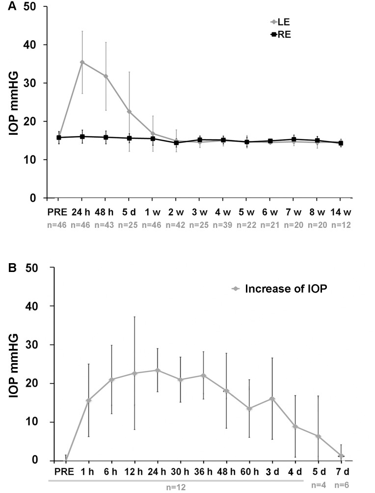
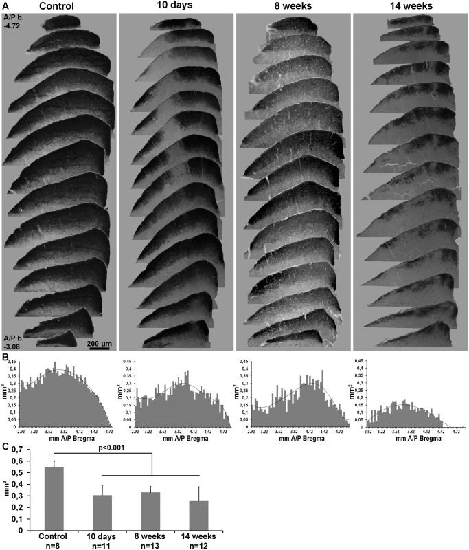
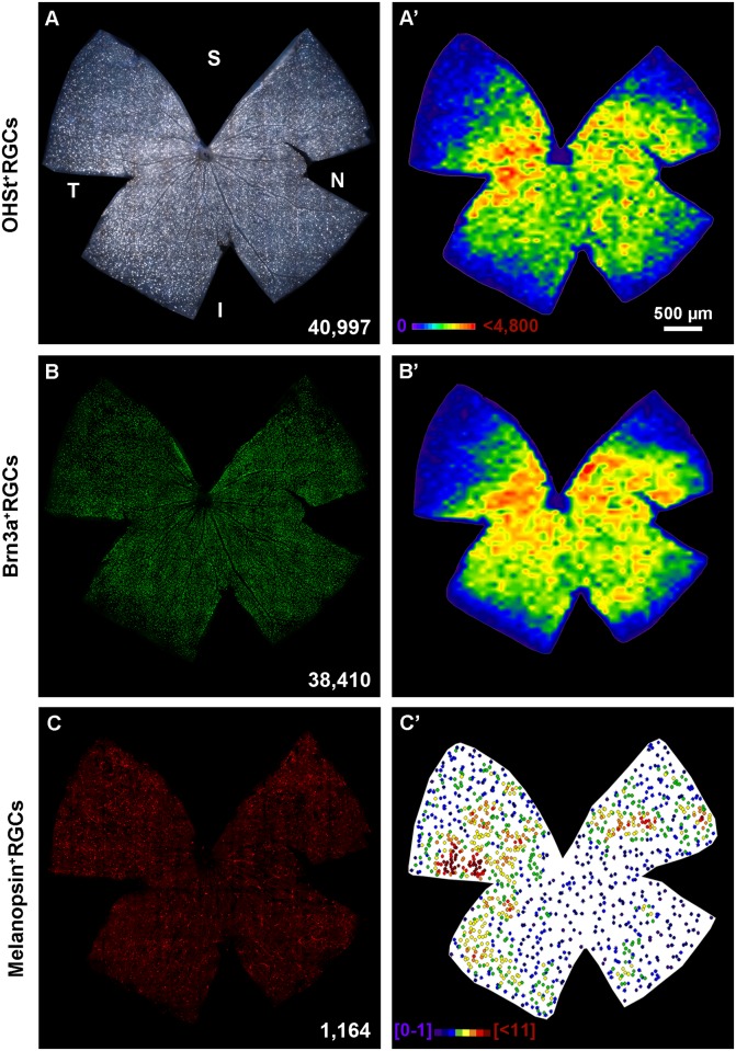
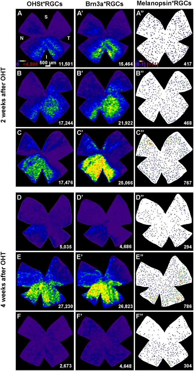
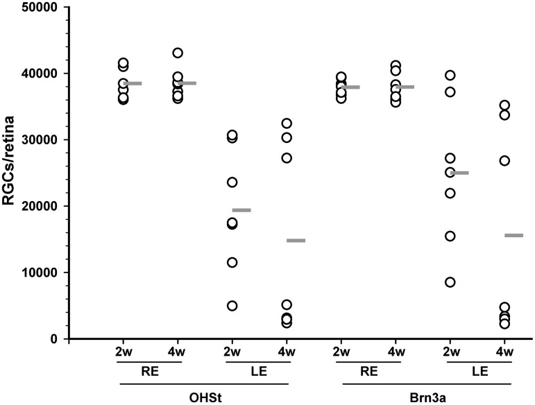
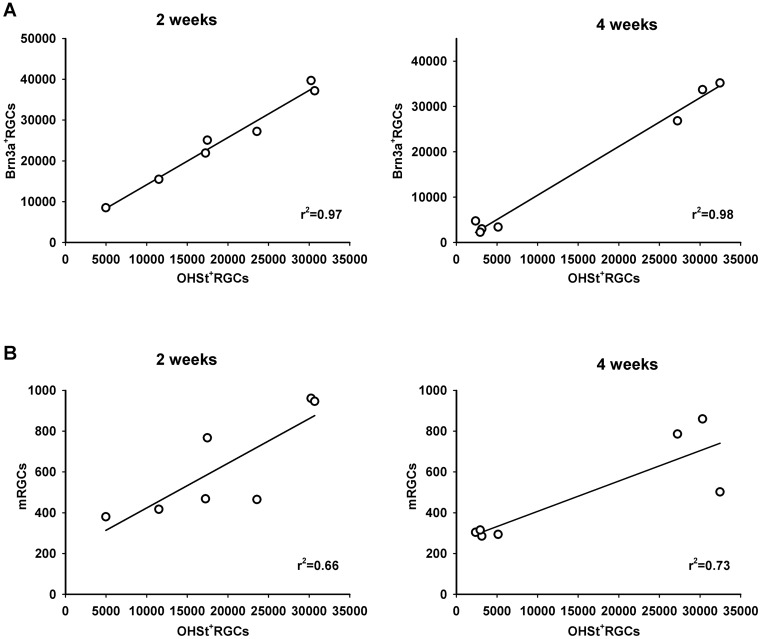
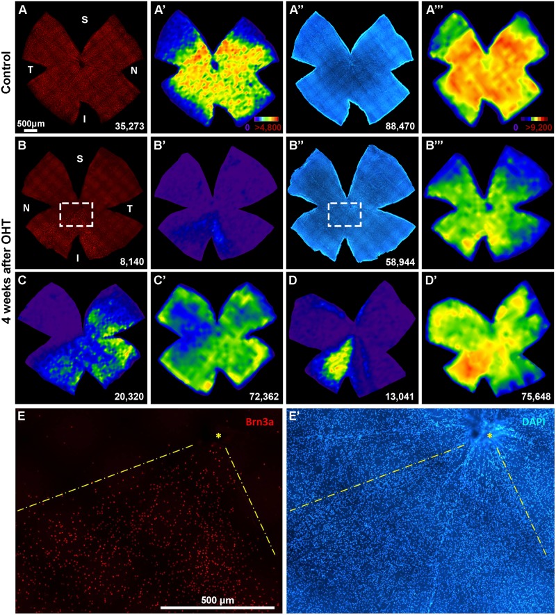
To study the effects of ocular hypertension (OHT) on the visual system of C57BL/6 pigmented mice, the limbal and episcleral veins of the left eye were laser photocoagulated (LP). LP increased the intraocular pressure during the first five days (d), reaching basal values at 7d. To investigate the effect of OHT on the retinal ganglion cell (RGC) retrograde axonal transport, hydroxistilbamidine methanesulfonate (OHSt) was applied to both superior colliculi (SCi) and the retinas were dissected 2 or 4 weeks after LP. To determine RGC survival, these same retinas were immunoreacted against Brn3a (general RGC population) and melanopsin (intrinsically photosensitive RGCs, m+RGCs). To study whether OHT affected non-RGC neurons in the ganglion cell layer (GCL), RGCs were immunodetected with Brn3a and all GCL nuclei counterstained with DAPI in a group of animals examined 4 weeks post-LP. Innervation of the SCi was examined at 10 days, 8 or 14 weeks after LP with the orthogradely transported cholera toxin subunit-B. OHT resulted in diffuse and sectorial loss of OHSt+RGCs (50% at 2 weeks and 62% at 4 weeks) and in a comparable loss of Brn3a+RGCs at the same time intervals. m+RGCs decreased to 59% at 2 weeks and to 46% at 4 weeks, such loss was diffuse, did not parallel the sectorial loss of the general RGC population and was more severe in the superior-temporal retina. In the GCL, cell loss is selective for RGCs and does not affect other non-RGC neurons. The retinotectal innervation appeared significantly reduced at 10 days (55.7%) and did not progress further up to 14 weeks (46.6%). Thus, LP-induced OHT results in retrograde degeneration of RGCs and m+RGCs, as well as in the loss of CTB-labelled retinotectal terminals.
为研究高眼压(OHT)对C57BL/6色素沉着小鼠视觉系统的影响,对左眼的角膜缘静脉和巩膜上静脉进行激光光凝(LP)。LP在最初五天(d)内使眼压升高,在第7天达到基础值。为研究OHT对视网膜神经节细胞(RGC)逆行轴突运输的影响,在LP后2周或4周,将甲磺酸羟乙磺脒(OHSt)应用于双侧上丘(SCi)并解剖视网膜。为确定RGC的存活情况,对这些相同的视网膜进行抗Brn3a(一般RGC群体)和黑视蛋白(内在光敏RGC,m+RGC)的免疫反应。为研究OHT是否影响神经节细胞层(GCL)中的非RGC神经元,在LP后4周检查的一组动物中,用Brn3a对RGC进行免疫检测,并用DAPI对所有GCL细胞核进行复染。在LP后10天、8周或14周,用正向运输的霍乱毒素亚基B检查SCi的神经支配情况。OHT导致OHSt+RGC弥漫性和扇形丢失(2周时为50%,4周时为62%),并在相同时间间隔内导致Brn3a+RGC出现类似程度的丢失。m+RGC在2周时降至59%,在4周时降至46%,这种丢失是弥漫性的,与一般RGC群体的扇形丢失不平行,并且在颞上视网膜中更严重。在GCL中,细胞丢失对RGC具有选择性,不影响其他非RGC神经元。视网膜顶盖神经支配在10天时显著减少(55.7%),直至14周时没有进一步进展(46.6%)。因此,LP诱导的OHT导致RGC和m+RGC逆行变性,以及CTB标记的视网膜顶盖终末丢失。