Zou Qihong, Yuan Bin-Ke, Gu Hong, Liu Dongqiang, Wang Danny J J, Gao Jia-Hong, Yang Yihong, Zang Yu-Feng
Center for MRI Research and Beijing City Key Lab for Medical Physics and Engineering, Peking University, Beijing, China; Neuroimaging Research Branch, National Institute on Drug Abuse, National Institutes of Health, Baltimore, Maryland, United States of America.
Center for Cognition and Brain Disorders, Affiliated Hospital, Hangzhou Normal University, Hangzhou, Zhejiang, China; Zhejiang Key Laboratory for Research in Assessment of Cognitive Impairments, Hangzhou Normal University, Hangzhou, Zhejiang, China.
PLoS One. 2015 Mar 27;10(3):e0121757. doi: 10.1371/journal.pone.0121757. eCollection 2015.
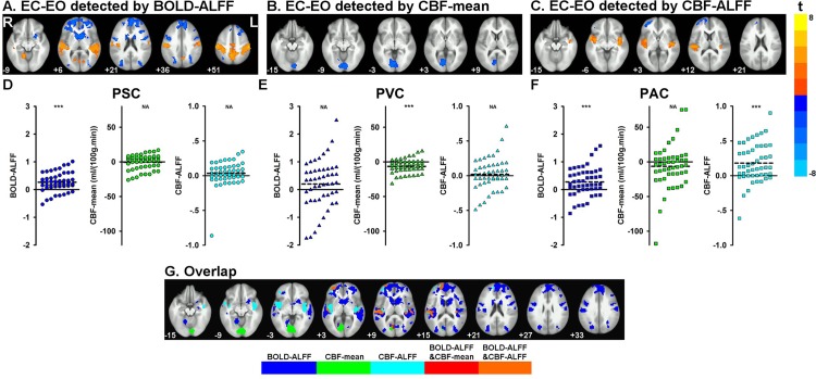
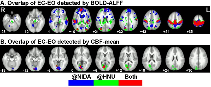
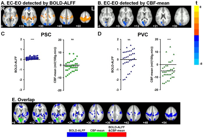
Resting-state fMRI studies have increasingly focused on multi-contrast techniques, such as BOLD and ASL imaging. However, these techniques may reveal different aspects of brain activity (e.g., static vs. dynamic), and little is known about the similarity or disparity of these techniques in detecting resting-state brain activity. It is therefore important to assess the static and dynamic characteristics of these fMRI techniques to guide future applications. Here we acquired fMRI data while subjects were in eyes-closed (EC) and eyes-open (EO) states, using both ASL and BOLD techniques, at two research centers (NIDA and HNU). Static brain activity was calculated as voxel-wise mean cerebral blood flow (CBF) using ASL, i.e., CBF-mean, while dynamic activity was measured by the amplitude of low frequency fluctuations (ALFF) of BOLD, i.e., BOLD-ALFF, at both NIDA and HNU, and CBF, i.e., CBF-ALFF, at NIDA. We showed that mean CBF was lower under EC than EO in the primary visual cortex, while BOLD-ALFF was higher under EC in the primary somatosensory cortices extending to the primary auditory cortices and lower in the lateral occipital area. Interestingly, mean CBF and BOLD-ALFF results overlapped at the visual cortex to a very small degree. Importantly, these findings were largely replicated by the HNU dataset. State differences found by CBF-ALFF were located in the primary auditory cortices, which were generally a subset of BOLD-ALFF and showed no spatial overlap with CBF-mean. In conclusion, static brain activity measured by mean CBF and dynamic brain activity measured by BOLD- and CBF-ALFF may reflect different aspects of resting-state brain activity and a combination of ASL and BOLD may provide complementary information on the biophysical and physiological processes of the brain.
静息态功能磁共振成像(fMRI)研究越来越多地聚焦于多对比技术,如血氧水平依赖(BOLD)成像和动脉自旋标记(ASL)成像。然而,这些技术可能揭示大脑活动的不同方面(例如,静态与动态),而对于这些技术在检测静息态大脑活动方面的相似性或差异知之甚少。因此,评估这些fMRI技术的静态和动态特征对于指导未来的应用很重要。在这里,我们在两个研究中心(美国国立药物滥用研究所和湖南大学)使用ASL和BOLD技术,在受试者闭眼(EC)和睁眼(EO)状态下采集了fMRI数据。在两个研究中心,静态大脑活动通过使用ASL计算体素水平的平均脑血流量(CBF)来确定,即CBF均值,而动态活动通过BOLD的低频波动幅度(ALFF)来测量,即BOLD-ALFF;在国立药物滥用研究所,动态活动还通过CBF的ALFF来测量,即CBF-ALFF。我们发现,在初级视觉皮层中,EC状态下的平均CBF低于EO状态,而在从初级体感皮层延伸到初级听觉皮层的区域,EC状态下的BOLD-ALFF较高,在枕叶外侧区域较低。有趣的是,平均CBF和BOLD-ALFF结果在视觉皮层的重叠程度非常小。重要的是,这些发现很大程度上被湖南大学的数据集所重复。CBF-ALFF发现的状态差异位于初级听觉皮层,这些区域通常是BOLD-ALFF的一部分,并且与CBF均值没有空间重叠。总之,通过平均CBF测量的静态大脑活动以及通过BOLD-ALFF和CBF-ALFF测量的动态大脑活动可能反映了静息态大脑活动的不同方面,并且ASL和BOLD的结合可能为大脑的生物物理和生理过程提供补充信息。