Suppr超能文献

基于微计算机断层扫描(μCT)对设计的聚对苯二甲酸丙二醇酯(PTMC)支架机械变形的评估。

μCT based assessment of mechanical deformation of designed PTMC scaffolds.

作者信息

Narra Nathaniel, Blanquer Sébastien B G, Haimi Suvi P, Grijpma Dirk W, Hyttinen Jari

机构信息

Department of Electronics and Communications Engineering, Tampere University of Technology, BioMediTech Tampere, Finland.

Department of Biomaterials Science and Technology, University of Twente, Enschede, The Netherlands.

出版信息

Clin Hemorheol Microcirc. 2015;60(1):99-108. doi: 10.3233/CH-151931.

Abstract

BACKGROUND

Advances in rapid-prototyping and 3D printing technologies have enhanced the possibilities in preparing designed architectures for tissue engineering applications. A major advantage in custom designing is the ability to create structures with desired mechanical properties. While the behaviour of a designed scaffold can be simulated using bulk material properties, it is important to verify the behaviour of a printed scaffold at the microstructure level.

OBJECTIVE

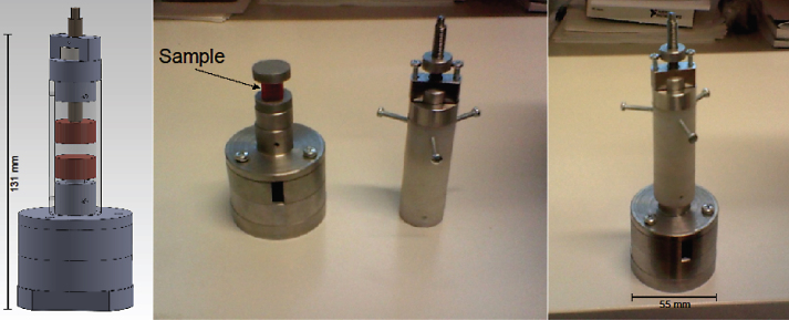
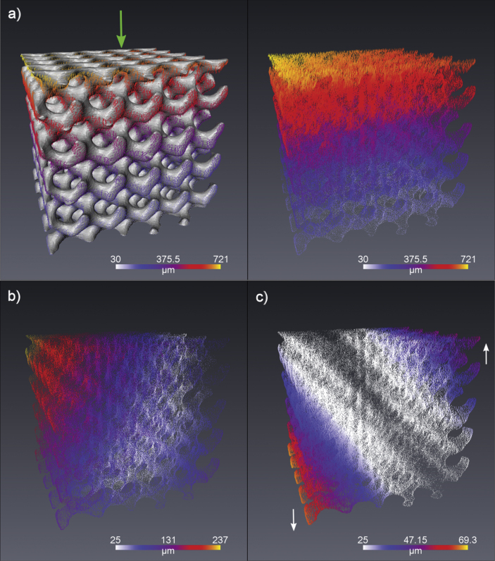
In this study we present an effective method in validating the mechanical behaviour of designed scaffolds using a μCT with an in-situ mechanical deformation device.

METHODS

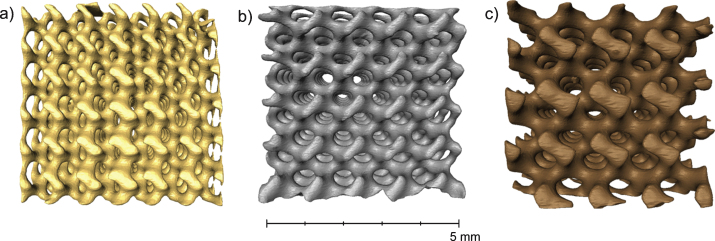
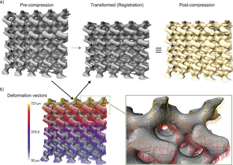
The scaffolds were prepared from biodegradable poly(trimethylene carbonate) (PTMC) by stereolithography and images obtained using a high-resolution μCT with 12.25μm isometric voxels. The data was processed (filtering, segmentation) and analysed (surface generation, registration) to extract relevant deformation features.

RESULTS

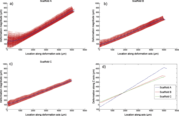
The computed local deformation fields, calculated at sub-pore resolutions, displayed expected linear behaviour within the scaffold along the compressions axis. On planes perpendicular to this axis, the deformations varied by 150- 200μm.

CONCLUSIONS

μCT based imaging with in-situ deformation provides a vital tool in validating the design parameters of printed scaffolds. Deformation fields obtained from micro-tomographic image volumes can serve to corroborate the simulated ideal design with the realized product.

摘要

背景

快速成型和3D打印技术的进步增加了为组织工程应用制备设计架构的可能性。定制设计的一个主要优势是能够创建具有所需机械性能的结构。虽然可以使用块状材料特性模拟设计支架的行为,但在微观结构层面验证打印支架的行为很重要。

目的

在本研究中,我们提出了一种使用带有原位机械变形装置的μCT验证设计支架机械行为的有效方法。

方法

通过立体光刻从可生物降解的聚碳酸三亚甲基酯(PTMC)制备支架,并使用具有12.25μm等距体素的高分辨率μCT获得图像。对数据进行处理(滤波、分割)和分析(表面生成、配准)以提取相关变形特征。

结果

在亚孔隙分辨率下计算的局部变形场在支架内沿压缩轴显示出预期的线性行为。在垂直于该轴的平面上,变形变化为150 - 200μm。

结论

基于μCT的原位变形成像为验证打印支架的设计参数提供了重要工具。从微观断层图像体积获得的变形场可用于将模拟的理想设计与实际产品进行对比。

https://cdn.ncbi.nlm.nih.gov/pmc/blobs/4808/4923732/c2ce084ccfa6/ch-60-1-ch1931-g001.jpg

相似文献

1
μCT based assessment of mechanical deformation of designed PTMC scaffolds.
Clin Hemorheol Microcirc. 2015;60(1):99-108. doi: 10.3233/CH-151931.
4
Triblock copolymers based on ε-caprolactone and trimethylene carbonate for the 3D printing of tissue engineering scaffolds.
Int J Artif Organs. 2017 May 9;40(4):176-184. doi: 10.5301/ijao.5000543. Epub 2017 Feb 1.
5
Preparation of a designed poly(trimethylene carbonate) microvascular network by stereolithography.
Adv Healthc Mater. 2014 Dec;3(12):2004-11. doi: 10.1002/adhm.201400363. Epub 2014 Oct 15.
6
Designing porosity and topography of poly(1,3-trimethylene carbonate) scaffolds.
Acta Biomater. 2009 Nov;5(9):3281-94. doi: 10.1016/j.actbio.2009.05.017. Epub 2009 May 20.
8
Flexible and elastic porous poly(trimethylene carbonate) structures for use in vascular tissue engineering.
Acta Biomater. 2010 Apr;6(4):1269-77. doi: 10.1016/j.actbio.2009.10.002. Epub 2009 Oct 7.
10
Preparation of Designed Poly(trimethylene carbonate) Meniscus Implants by Stereolithography: Challenges in Stereolithography.
Macromol Biosci. 2016 Dec;16(12):1853-1863. doi: 10.1002/mabi.201600290. Epub 2016 Oct 17.

引用本文的文献

2
Analysis of 3D Prints by X-ray Computed Microtomography and Terahertz Pulsed Imaging.
Pharm Res. 2017 May;34(5):1037-1052. doi: 10.1007/s11095-016-2083-1. Epub 2016 Dec 21.

本文引用的文献

2
Current trends in the design of scaffolds for computer-aided tissue engineering.
Acta Biomater. 2014 Feb;10(2):580-94. doi: 10.1016/j.actbio.2013.10.024. Epub 2013 Oct 30.
3
Computation of full-field displacements in a scaffold implant using digital volume correlation and finite element analysis.
Med Eng Phys. 2013 Sep;35(9):1298-312. doi: 10.1016/j.medengphy.2013.02.001. Epub 2013 Mar 6.
4
Manipulating failure mechanism of rapid prototyped scaffolds by changing nodal connectivity and geometry of the pores.
J Biomech. 2012 Nov 15;45(16):2866-75. doi: 10.1016/j.jbiomech.2012.08.029. Epub 2012 Sep 15.
5
Preparation of flexible and elastic poly(trimethylene carbonate) structures by stereolithography.
Macromol Biosci. 2011 Dec 8;11(12):1662-71. doi: 10.1002/mabi.201100203. Epub 2011 Oct 17.
6
Mathematically defined tissue engineering scaffold architectures prepared by stereolithography.
Biomaterials. 2010 Sep;31(27):6909-16. doi: 10.1016/j.biomaterials.2010.05.068. Epub 2010 Jun 26.
7
A review on stereolithography and its applications in biomedical engineering.
Biomaterials. 2010 Aug;31(24):6121-30. doi: 10.1016/j.biomaterials.2010.04.050. Epub 2010 May 15.
8
On the biomechanical function of scaffolds for engineering load-bearing soft tissues.
Acta Biomater. 2010 Jul;6(7):2365-81. doi: 10.1016/j.actbio.2010.01.001. Epub 2010 Jan 7.
9
Cell-scaffold mechanical interplay within engineered tissue.
Semin Cell Dev Biol. 2009 Aug;20(6):656-64. doi: 10.1016/j.semcdb.2009.02.001. Epub 2009 Feb 13.
10
Accuracy and precision of digital volume correlation in quantifying displacements and strains in trabecular bone.
J Biomech. 2007;40(15):3516-20. doi: 10.1016/j.jbiomech.2007.04.019. Epub 2007 Jun 13.

文献AI研究员

20分钟写一篇综述,助力文献阅读效率提升50倍。

立即体验

用中文搜PubMed

大模型驱动的PubMed中文搜索引擎

马上搜索

文档翻译

学术文献翻译模型,支持多种主流文档格式。

立即体验