Fausther Michel, Goree Jessica R, Lavoie Élise G, Graham Alicia L, Sévigny Jean, Dranoff Jonathan A
Division of Gastroenterology & Hepatology, University of Arkansas for Medical Sciences, Little Rock, AR, United States of America; Research Service, Central Arkansas VA Healthcare System, Little Rock, AR, United States of America.
Division of Gastroenterology & Hepatology, University of Arkansas for Medical Sciences, Little Rock, AR, United States of America.
PLoS One. 2015 Mar 30;10(3):e0121161. doi: 10.1371/journal.pone.0121161. eCollection 2015.
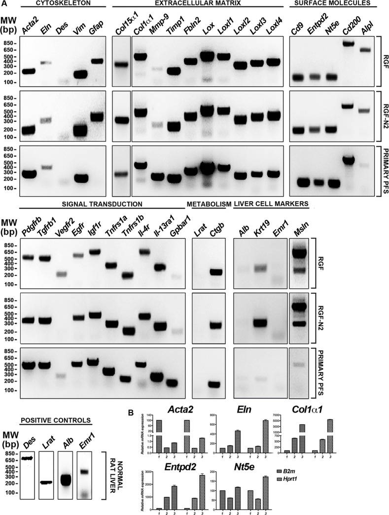
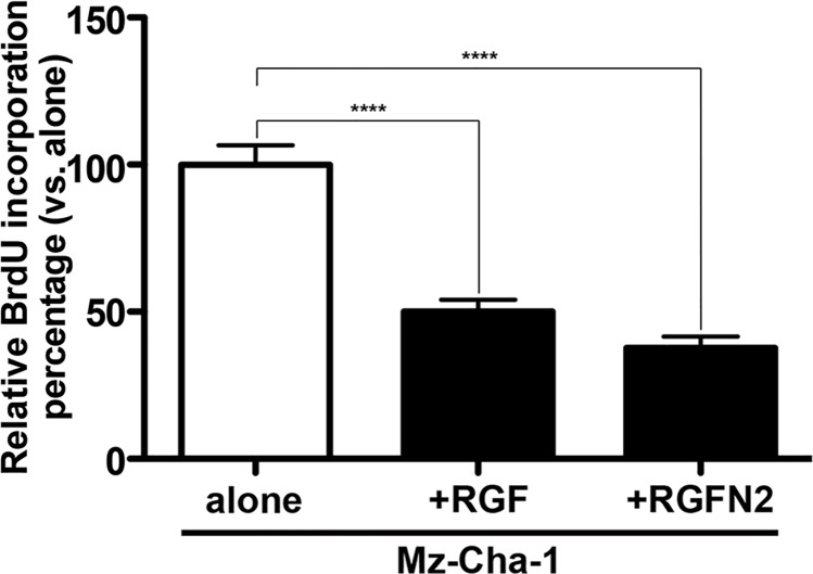
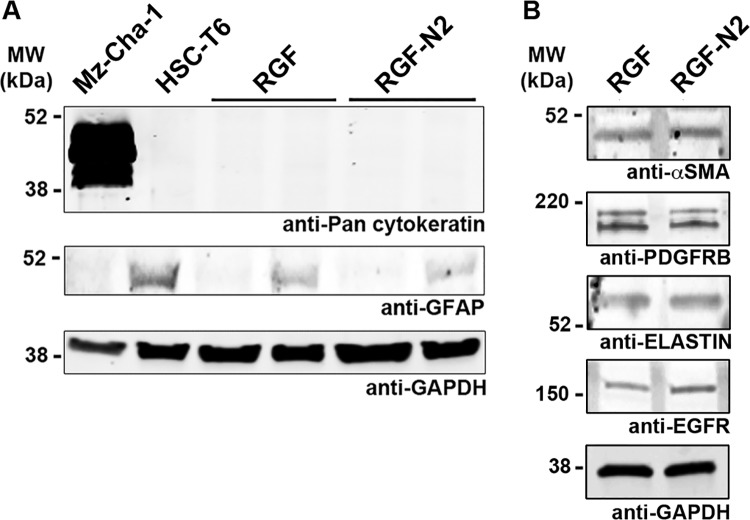
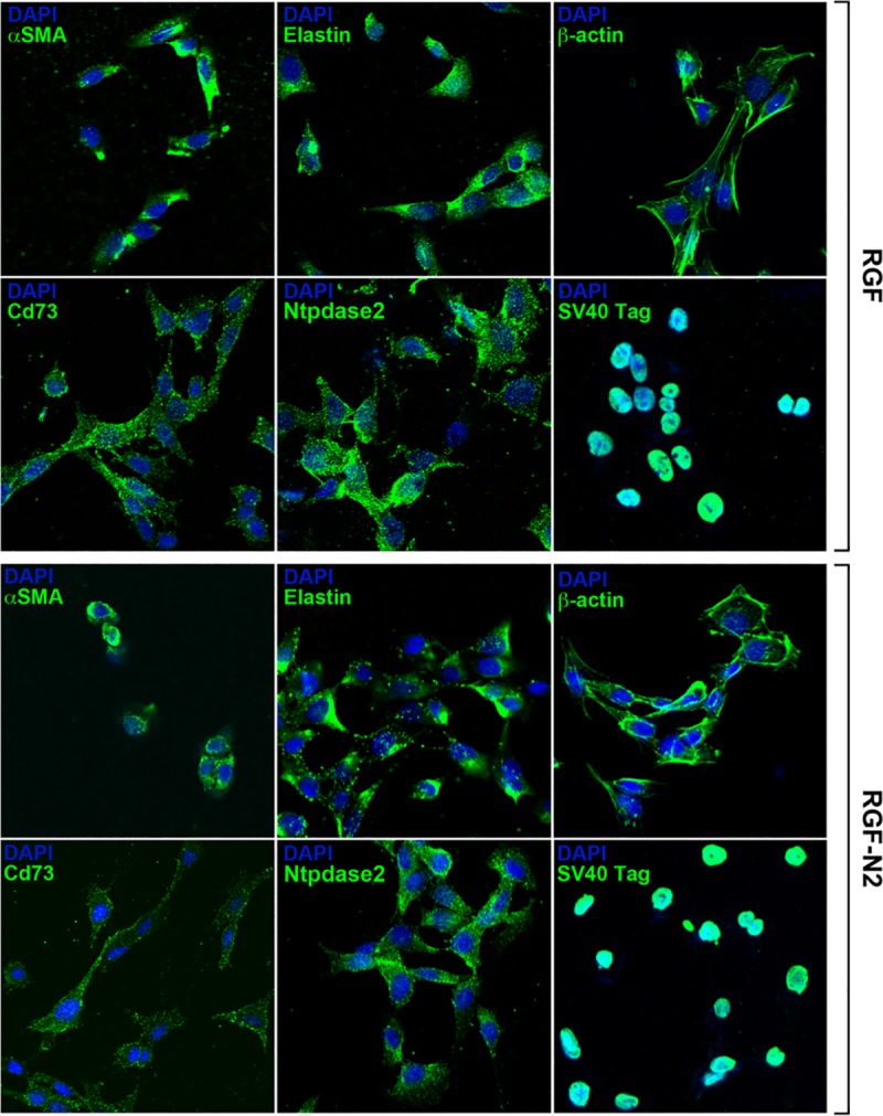
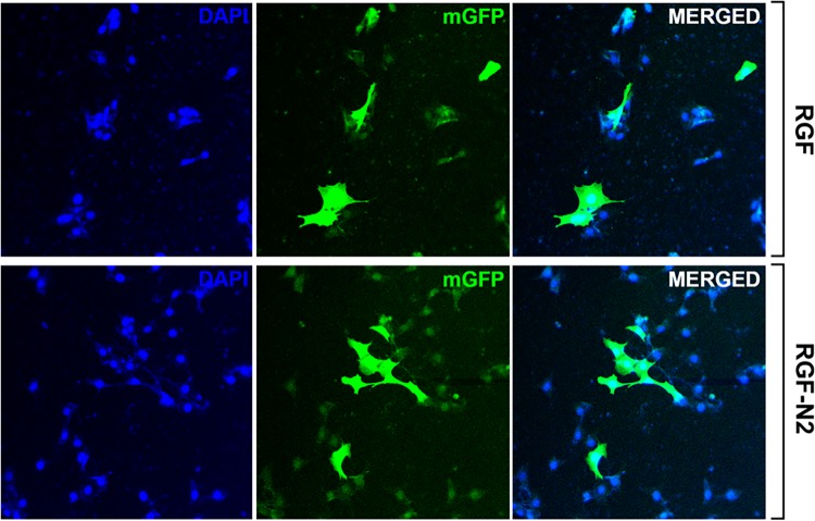
The major sources of scar-forming myofibroblasts during liver fibrosis are activated hepatic stellate cells (HSC) and portal fibroblasts (PF). In contrast to well-characterized HSC, PF remain understudied and poorly defined. This is largely due to the facts that isolation of rodent PF for functional studies is technically challenging and that PF cell lines had not been established. To address this, we have generated two polyclonal portal myofibroblast cell lines, RGF and RGF-N2. RGF and RGF-N2 were established from primary PF isolated from adult rat livers that underwent culture activation and subsequent SV40-mediated immortalization. Specifically, Ntpdase2/Cd39l1-sorted primary PF were used to generate the RGF-N2 cell line. Both cell lines were functionally characterized by RT-PCR, immunofluorescence, immunoblot and bromodeoxyuridine-based proliferation assay. First, immortalized RGF and RGF-N2 cells are positive for phenotypic myofibroblast markers alpha smooth muscle actin, type I collagen alpha-1, tissue inhibitor of metalloproteinases-1, PF-specific markers elastin, type XV collagen alpha-1 and Ntpdase2/Cd39l1, and mesenchymal cell marker ecto-5'-nucleotidase/Cd73, while negative for HSC-specific markers desmin and lecithin retinol acyltransferase. Second, both RGF and RGF-N2 cell lines are readily transfectable using standard methods. Finally, RGF and RGF-N2 cells attenuate the growth of Mz-ChA-1 cholangiocarcinoma cells in co-culture, as previously demonstrated for primary PF. Immortalized rat portal myofibroblast RGF and RGF-N2 cell lines express typical markers of activated PF-derived myofibroblasts, are suitable for DNA transfection, and can effectively inhibit cholangiocyte proliferation. Both RGF and RGF-N2 cell lines represent novel in vitro cellular models for the functional studies of portal (myo)fibroblasts and their contribution to the progression of liver fibrosis.
肝纤维化过程中形成瘢痕的肌成纤维细胞的主要来源是活化的肝星状细胞(HSC)和门周成纤维细胞(PF)。与特征明确的HSC不同,PF的研究较少且定义不明确。这主要是由于以下事实:分离啮齿动物PF用于功能研究在技术上具有挑战性,并且尚未建立PF细胞系。为了解决这个问题,我们生成了两种多克隆门周肌成纤维细胞系,RGF和RGF-N2。RGF和RGF-N2是从成年大鼠肝脏分离的原代PF中建立的,这些原代PF经过培养活化和随后的SV40介导的永生化。具体而言,使用Ntpdase2/Cd39l1分选的原代PF来生成RGF-N2细胞系。通过逆转录聚合酶链反应(RT-PCR)、免疫荧光、免疫印迹和基于溴脱氧尿苷的增殖试验对这两种细胞系进行了功能表征。首先,永生化的RGF和RGF-N2细胞对于表型肌成纤维细胞标志物α平滑肌肌动蛋白、I型胶原α-1、金属蛋白酶组织抑制剂-1、PF特异性标志物弹性蛋白、XV型胶原α-1和Ntpdase2/Cd39l1以及间充质细胞标志物外5'-核苷酸酶/Cd73呈阳性,而对于HSC特异性标志物结蛋白和卵磷脂视黄醇酰基转移酶呈阴性。其次,RGF和RGF-N2细胞系都可以使用标准方法轻松转染。最后,RGF和RGF-N2细胞在共培养中可减弱Mz-ChA-1胆管癌细胞的生长,正如之前原代PF所显示的那样。永生化的大鼠门周肌成纤维细胞RGF和RGF-N2细胞系表达活化的PF衍生肌成纤维细胞的典型标志物,适用于DNA转染,并且可以有效抑制胆管细胞增殖。RGF和RGF-N2细胞系均代表用于门周(肌)成纤维细胞功能研究及其对肝纤维化进展贡献的新型体外细胞模型。