Johnson Errin, Seiradake Elena, Jones E Yvonne, Davis Ilan, Grünewald Kay, Kaufmann Rainer
Sir William Dunn School of Pathology, University of Oxford, South Parks Road, Oxford, OX1 3RE, UK.
Division of Structural Biology, Wellcome Trust Centre for Human Genetics, University of Oxford, Roosevelt Drive, Oxford, OX3 7BN, UK.
Sci Rep. 2015 Mar 31;5:9583. doi: 10.1038/srep09583.
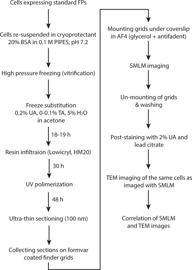
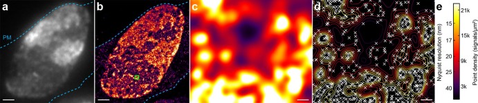
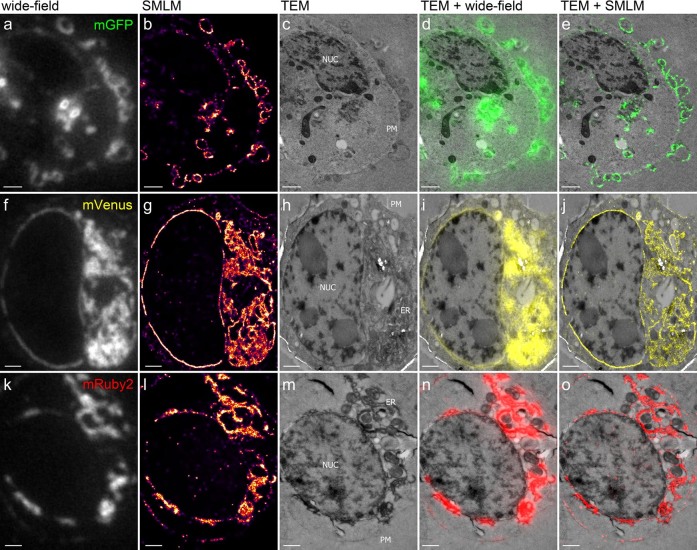
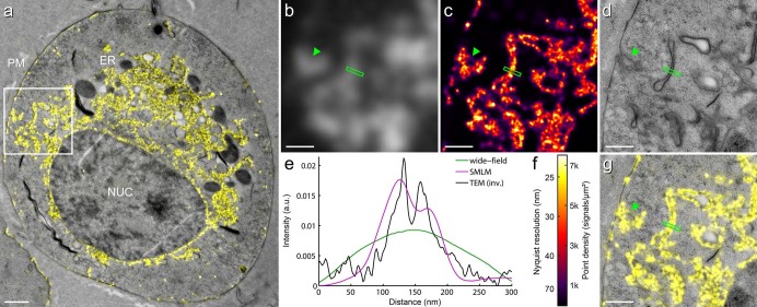
We introduce a method for correlative in-resin super-resolution fluorescence and electron microscopy (EM) of biological structures in mammalian culture cells. Cryo-fixed resin embedded samples offer superior structural preservation, performing in-resin super-resolution, however, remains a challenge. We identified key aspects of the sample preparation procedure of high pressure freezing, freeze substitution and resin embedding that are critical for preserving fluorescence and photo-switching of standard fluorescent proteins, such as mGFP, mVenus and mRuby2. This enabled us to combine single molecule localization microscopy with transmission electron microscopy imaging of standard fluorescent proteins in cryo-fixed resin embedded cells. We achieved a structural resolution of 40-50 nm (~17 nm average single molecule localization accuracy) in the fluorescence images without the use of chemical fixation or special fluorophores. Using this approach enabled the correlation of fluorescently labeled structures to the ultrastructure in the same cell at the nanometer level and superior structural preservation.
我们介绍了一种用于对哺乳动物培养细胞中的生物结构进行树脂内超分辨率荧光与电子显微镜(EM)关联成像的方法。冷冻固定的树脂包埋样品能提供卓越的结构保存效果,然而,进行树脂内超分辨率成像仍然是一项挑战。我们确定了高压冷冻、冷冻置换和树脂包埋样品制备过程中的关键环节,这些环节对于保留标准荧光蛋白(如mGFP、mVenus和mRuby2)的荧光和光开关特性至关重要。这使我们能够将单分子定位显微镜与冷冻固定树脂包埋细胞中标准荧光蛋白的透射电子显微镜成像相结合。在不使用化学固定或特殊荧光团的情况下,我们在荧光图像中实现了40 - 50纳米的结构分辨率(平均单分子定位精度约为17纳米)。使用这种方法能够在纳米水平上对同一细胞中荧光标记结构与超微结构进行关联,并实现卓越的结构保存。