College of Electronic and Information Engineering of Hebei University, Baoding, 071002 China.
Health Inf Sci Syst. 2013 Jan 10;1:5. doi: 10.1186/2047-2501-1-5. eCollection 2013.
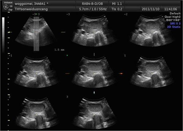
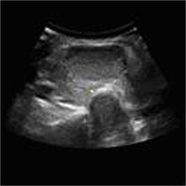
The incidence of thyroid nodule is very high and generally increases with the age. Thyroid nodule may presage the emergence of thyroid cancer. Most thyroid nodules are asymptomatic which makes thyroid cancer different from other cancers. The thyroid nodule can be completely cured if detected early. Therefore, it is necessary to correctly classify the thyroid nodule to be benign or malignant. Fine needle aspiration cytology is a recognized early diagnosis method of thyroid nodule. There are still some limitations in the fine needle aspiration cytology, such as the difficulty in location and the insufficient cytology specimen. The accuracy of ultrasound diagnosis of thyroid nodule improves constantly, and it has become the first choice for auxiliary examination of thyroid nodular disease. If we could combine medical imaging technology and fine needle aspiration cytology, the diagnostic rate of thyroid nodule would be improved significantly. The properties of ultrasound, such as echo, shadow, and reflection, will degrade the image quality, which makes it difficult to recognize the edges for physicians. Image segmentation technique based on graph theory has become a research hotspot at present. Normalized cut (Ncut) is a representative one, whose biggest advantage is not prone to small region segmentation but suitable for segmentation of feature parts of medical image. However, how to solve the normalized cut has become a problem, which needs large memory capacity and heavy calculation of weight matrix. It always generates over segmentation or less segmentation which leads to inaccurate in the segmentation. The speckle noise produced in the formation process of B ultrasound image of thyroid tumor makes the quality of the image deteriorate. In the light of this characteristic, we combine the anisotropic diffusion model with the normalized cut in this paper. After the enhancement of anisotropic diffusion model, it removes the noise in the B ultrasound image while preserves the important edges and local details. This reduces the amount of computation in constructing the weight matrix of the improved normalized cut and improves the accuracy of the final segmentation results. The feasibility of the method is proved by the experimental results.
甲状腺结节的发病率非常高,且通常随年龄增长而增加。甲状腺结节可能预示着甲状腺癌的出现。大多数甲状腺结节无症状,这使甲状腺癌有别于其他癌症。如果早期发现,甲状腺结节可以完全治愈。因此,有必要正确分类甲状腺结节的良恶性。细针穿刺细胞学检查是一种公认的甲状腺结节早期诊断方法。然而,细针穿刺细胞学检查仍存在一些局限性,例如定位困难和细胞学标本不足。甲状腺结节的超声诊断准确性不断提高,已成为甲状腺结节疾病辅助检查的首选。如果能将医学影像学技术与细针穿刺细胞学检查相结合,将显著提高甲状腺结节的诊断率。超声的回声、阴影和反射等特性会降低图像质量,使医生难以识别边缘。基于图论的图像分割技术目前已成为研究热点。归一化割(Ncut)是一种有代表性的方法,其最大的优势是不易出现小区域分割,而是适用于医学图像特征部分的分割。然而,如何解决归一化割问题已成为一个难题,因为它需要大量的内存容量和沉重的权重矩阵计算。这往往会导致过度分割或欠分割,从而导致分割不准确。甲状腺肿瘤 B 超图像在形成过程中产生的斑点噪声会使图像质量恶化。针对这一特点,本文将各向异性扩散模型与归一化割相结合。各向异性扩散模型增强后,在去除 B 超图像中噪声的同时保留了重要的边缘和局部细节,减少了改进的归一化割中构建权重矩阵的计算量,提高了最终分割结果的准确性。实验结果证明了该方法的可行性。