Jin Juebin, Zhu Haiyan, Zhang Jindi, Ai Yao, Zhang Ji, Teng Yinyan, Xie Congying, Jin Xiance
Department of Medical Engineering, Wenzhou Medical University First Affiliated Hospital, Wenzhou, China.
Department of Gynecology, Shanghai First Maternal and Infant Hospital, Tongji University School of Medicine, Shanghai, China.
Front Oncol. 2021 Feb 18;10:614201. doi: 10.3389/fonc.2020.614201. eCollection 2020.
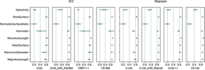
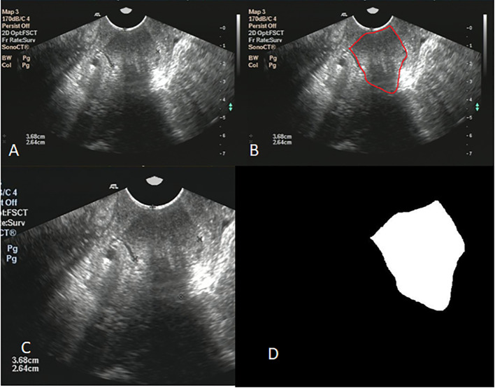
Few studies have reported the reproducibility and stability of ultrasound (US) images based radiomics features obtained from automatic segmentation in oncology. The purpose of this study is to study the accuracy of automatic segmentation algorithms based on multiple U-net models and their effects on radiomics features from US images for patients with ovarian cancer. A total of 469 US images from 127 patients were collected and randomly divided into three groups: training sets (353 images), validation sets (23 images), and test sets (93 images) for automatic segmentation models building. Manual segmentation of target volumes was delineated as ground truth. Automatic segmentations were conducted with U-net, U-net++, U-net with Resnet as the backbone (U-net with Resnet), and CE-Net. A python 3.7.0 and package Pyradiomics 2.2.0 were used to extract radiomic features from the segmented target volumes. The accuracy of automatic segmentations was evaluated by Jaccard similarity coefficient (JSC), dice similarity coefficient (DSC), and average surface distance (ASD). The reliability of radiomics features were evaluated by Pearson correlation and intraclass correlation coefficients (ICC). CE-Net and U-net with Resnet outperformed U-net and U-net++ in accuracy performance by achieving a DSC, JSC, and ASD of 0.87, 0.79, 8.54, and 0.86, 0.78, 10.00, respectively. A total of 97 features were extracted from the delineated target volumes. The average Pearson correlation was 0.86 (95% CI, 0.83-0.89), 0.87 (95% CI, 0.84-0.90), 0.88 (95% CI, 0.86-0.91), and 0.90 (95% CI, 0.88-0.92) for U-net++, U-net, U-net with Resnet, and CE-Net, respectively. The average ICC was 0.84 (95% CI, 0.81-0.87), 0.85 (95% CI, 0.82-0.88), 0.88 (95% CI, 0.85-0.90), and 0.89 (95% CI, 0.86-0.91) for U-net++, U-net, U-net with Resnet, and CE-Net, respectively. CE-Net based segmentation achieved the best radiomics reliability. In conclusion, U-net based automatic segmentation was accurate enough to delineate the target volumes on US images for patients with ovarian cancer. Radiomics features extracted from automatic segmented targets showed good reproducibility and for reliability further radiomics investigations.
很少有研究报告过从肿瘤学中的自动分割获得的基于超声(US)图像的放射组学特征的可重复性和稳定性。本研究的目的是研究基于多个U-net模型的自动分割算法的准确性及其对卵巢癌患者US图像放射组学特征的影响。共收集了127例患者的469幅US图像,并随机分为三组:训练集(353幅图像)、验证集(23幅图像)和测试集(93幅图像),用于构建自动分割模型。将目标体积的手动分割划定为真实情况。使用U-net、U-net++、以Resnet为骨干的U-net(U-net with Resnet)和CE-Net进行自动分割。使用python 3.7.0和Pyradiomics 2.2.0软件包从分割后的目标体积中提取放射组学特征。通过杰卡德相似系数(JSC)、骰子相似系数(DSC)和平均表面距离(ASD)评估自动分割的准确性。通过皮尔逊相关性和组内相关系数(ICC)评估放射组学特征的可靠性。CE-Net和U-net with Resnet在准确性方面优于U-net和U-net++,其DSC、JSC和ASD分别为0.87、0.79和8.54,以及0.86、0.78和10.00。从划定的目标体积中总共提取了97个特征。U-net++、U-net、U-net with Resnet和CE-Net的平均皮尔逊相关性分别为0.86(95%CI,0.83-0.89)、0.87(95%CI,0.84-0.90)、0.88(95%CI,0.86-。91)和0.90(95%CI,0.88-0.92)。U-net++、U-net、U-net with Resnet和CE-Net的平均ICC分别为0.84(95%CI,0.81-0.87)、0.85(95%CI,0.82-0.88)、0.88(95%CI,0.85-0.90)和0.89(95%CI,0.86-0.91)。基于CE-Net的分割实现了最佳的放射组学可靠性。总之,基于U-net的自动分割对于勾勒卵巢癌患者US图像上的目标体积足够准确。从自动分割目标中提取的放射组学特征显示出良好的可重复性,为进一步的放射组学研究提供了可靠性。