Dokainish Hisham
Glob Cardiol Sci Pract. 2015 Jan 26;2015:3. doi: 10.5339/gcsp.2015.3. eCollection 2015.
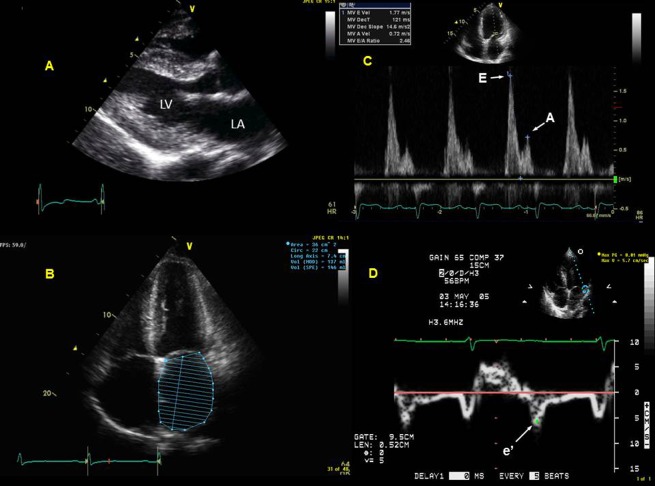
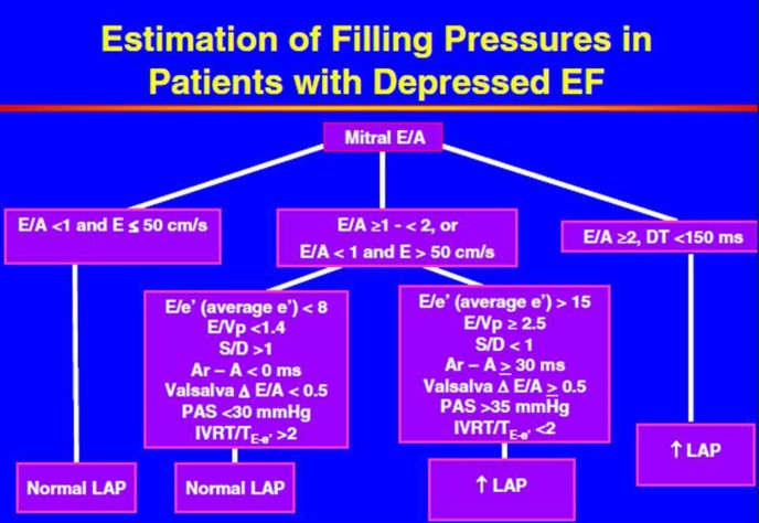
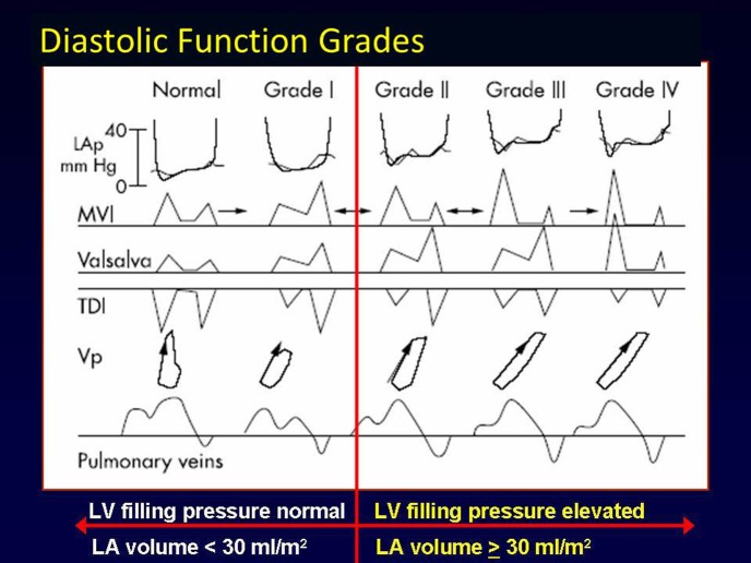
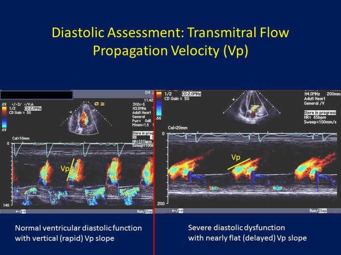
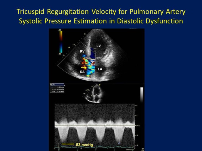
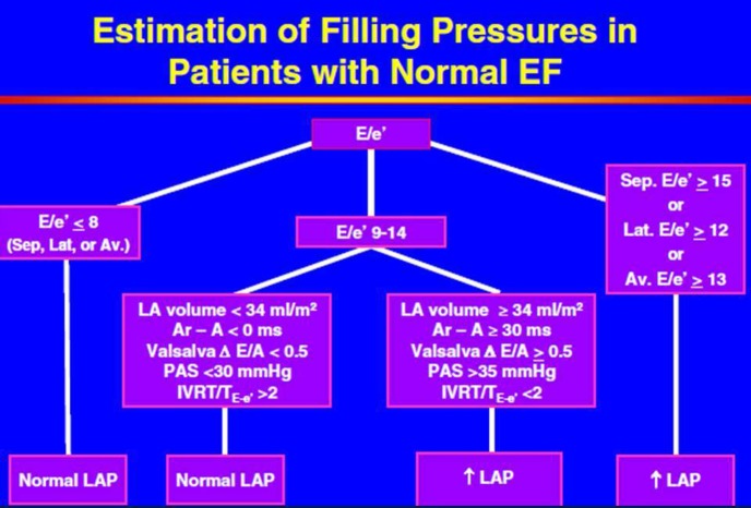
Comprehensive and precise assessment of left ventricular (LV) systolic and diastolic function is necessary to establish, or exclude, heart failure as a cause or component of dyspnea. Echocardiography with Doppler readily assesses LV diastolic function; advantages include that echocardiography is non-invasive, does not require radiation, is portable, rapid, readily available, and in competent hands, can provide an accurate and comprehensive assessment of LV systolic and diastolic function. Correct assessment of LV diastolic function is relevant in patients with both depressed and preserved LV ejection fraction (EF ≥ 50%, and < 50%, respectively). Tissue Doppler (TD) imaging has been useful in demonstrating impaired LV relaxation in the setting of preserved LVEF, which, in the setting of increased cardiac volume, can result in elevated LV filling pressures, and dyspnea due to diastolic heart failure. TD imaging is not always critical in patients with depressed LVEF, since such patients by definition have impaired LV relaxation, and thus significant increases in volume will result in increases in LV filling pressure due to impaired LV compliance. Thus, in depressed LVEF, transmitral flow velocities (E and A, and E/A) and deceleration time, pulmonary venous Doppler, left atrial volume, and pulmonary artery (PA) pressures suffice for the accurate assessment of LV filling pressures. Overall, diastolic assessment by echo-Doppler can be readily achieved in by using a comprehensive diastolic assessment-incorporating many 2-dimensional, conventional and tissue Doppler variables-as opposed to relying on any single, diastolic parameter, which can lead to errors.
对左心室(LV)收缩和舒张功能进行全面而精确的评估,对于确定或排除心力衰竭作为呼吸困难的病因或组成部分至关重要。采用多普勒的超声心动图能够轻松评估左心室舒张功能;其优点包括超声心动图是非侵入性的,无需辐射,便于携带,操作快速,随时可用,并且在技术熟练的人员操作下,能够对左心室收缩和舒张功能进行准确而全面的评估。左心室舒张功能的正确评估对于左心室射血分数(EF)降低和保留(分别为EF≥50%和<50%)的患者均具有重要意义。组织多普勒(TD)成像有助于在左心室射血分数保留的情况下显示左心室舒张功能受损,在心脏容量增加的情况下,这可能导致左心室充盈压升高以及舒张性心力衰竭引起的呼吸困难。在左心室射血分数降低的患者中,TD成像并非总是至关重要,因为根据定义,此类患者左心室舒张功能受损,因此容量的显著增加将由于左心室顺应性受损而导致左心室充盈压升高。因此,在左心室射血分数降低的情况下,二尖瓣血流速度(E和A以及E/A)、减速时间、肺静脉多普勒、左心房容积和肺动脉(PA)压力足以准确评估左心室充盈压。总体而言,通过使用包含许多二维、传统和组织多普勒变量的全面舒张功能评估,而不是依赖任何单一的舒张参数,超声多普勒舒张功能评估可以很容易地实现,因为单一参数可能导致错误。