Rodionov R, Bartlett P A, He Ci, Vos S B, Focke N K, Ourselin S G, Duncan J S
Department of Clinical and Experimental Epilepsy, UCL Institute of Neurology, London, UK ; MRI Unit, Epilepsy Society, Chalfont St Peter, Buckinghamshire, UK.
Department of Clinical and Experimental Epilepsy, UCL Institute of Neurology, London, UK ; MRI Unit, Epilepsy Society, Chalfont St Peter, Buckinghamshire, UK ; Department of Radiology, Chengdu Military General Hospital, China.
Neuroimage Clin. 2015 Mar 13;7:788-91. doi: 10.1016/j.nicl.2015.03.004. eCollection 2015.
Qualitatively, FLAIR MR imaging is sensitive to the detection of hippocampal sclerosis (HS). Quantitative analysis of T2 maps provides a useful objective measure and increased sensitivity over visual inspection of T2-weighted scans. We aimed to determine whether quantification of normalised FLAIR is as sensitive as T2 mapping in detection of HS.
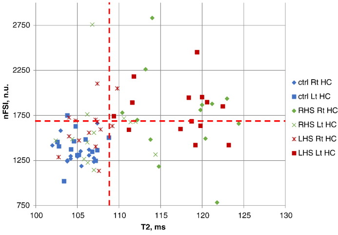
Dual echo T2 and FLAIR MR images were retrospectively analysed in 27 patients with histologically confirmed HS and increased T2 signal in ipsilateral hippocampus and 14 healthy controls. Regions of interest were manually segmented in all hippocampi aiming to avoid inclusion of CSF. Hippocampal T2 values and measures of normalised FLAIR Signal Intensity (nFSI) were compared in healthy and sclerotic hippocampi.
HS was identified on T2 values with 100% sensitivity and 100% specificity. HS was identified on nFSI measures with 60% sensitivity and 93% specificity.
T2 mapping is superior to nFSI for identification of HS.
定性地说,液体衰减反转恢复(FLAIR)磁共振成像(MRI)对检测海马硬化(HS)很敏感。T2图的定量分析提供了一种有用的客观测量方法,并且比T2加权扫描的视觉检查具有更高的敏感性。我们旨在确定标准化FLAIR的定量分析在检测HS方面是否与T2成像一样敏感。
回顾性分析了27例经组织学证实为HS且同侧海马T2信号增强的患者以及14名健康对照者的双回波T2和FLAIR MR图像。在所有海马中手动分割感兴趣区域,以避免包含脑脊液。比较了健康海马和硬化海马的海马T2值和标准化FLAIR信号强度(nFSI)测量值。
基于T2值识别HS的敏感性为100%,特异性为100%。基于nFSI测量值识别HS的敏感性为60%,特异性为93%。
在识别HS方面,T2成像优于nFSI。