Department of Neurosurgery, Peking Union Medical College Hospital, Beijing, China.
BrainNow Research Institute, Shenzhen, Guangdong Province, China.
BMC Med Imaging. 2020 Apr 25;20(1):42. doi: 10.1186/s12880-020-00440-z.
Hippocampal sclerosis (HS) is associated with post-surgery outcome in patients with temporal lobe epilepsy (TLE), and an automated method that quantifies HS severity is still lacking. Here, we aim to propose an MRI-based HS index (HSI) that integrates hippocampal volume and FLAIR signal to measure the severity of HS.
Forty-two pre-surgery TLE patients were included retrospectively, with T1-weighted (T1W) and FLAIR images acquired from each subject. Two experienced neurosurgeons (W.D. and C.S.) and one neurologist (Q.L.) rated HS severity with a four-class grading scale (normal, mild, moderate and severe) based on both hippocampal volume loss and increased FLAIR signal. A consensus of HS severity for each subject was made by voting among the three visual rating results. Regarding the automatic quantification, the hippocampal volume was quantified by AccuBrain on T1W image, and the FLAIR signal of hippocampus was calculated as the mean intensity of hippocampal region on the FLAIR image (normalized by the mean intensity of gray matter). To fit the HSI from visual rating, we applied ordinal regression with the voted visual rating as the dependent variable, and hippocampal volume and FLAIR signal as the independent variables. The HSI was calculated by weighting the predicted probabilities of the four-class grading scales from ordinal regression.
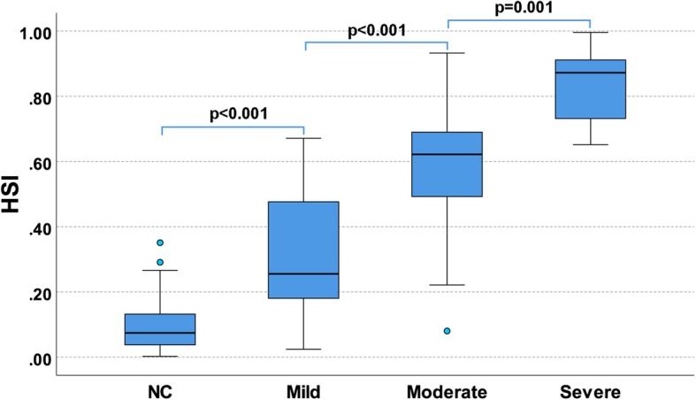
The intra-class correlation coefficient (single measure) of the three raters was 0.806. The generated HSI was significantly correlated with the visual rating scales of the three raters (W.D.: 0.823, Q.L.: 0.817, C.S.: 0.717). HSI scores well differentiated the different HS categories as defined by the agreed HS visual rating (normal vs. mild: p < 0.001, mild vs. moderate: p < 0.001, moderate vs. severe: p = 0.001).
The proposed HSI was consistent with visual rating scales from epileptologists and sensitive to HS severity. This MRI-based index may help to evaluate HS severity in clinical practice. Further validations are needed to associate HSI with post-surgery outcomes.
海马硬化(HS)与颞叶癫痫(TLE)患者手术后的结果有关,目前仍然缺乏一种能够自动量化 HS 严重程度的方法。本研究旨在提出一种基于 MRI 的 HS 指数(HSI),该指数综合了海马体积和 FLAIR 信号,用于测量 HS 的严重程度。
回顾性纳入 42 例术前 TLE 患者,每位患者均采集 T1 加权(T1W)和 FLAIR 图像。由 2 名经验丰富的神经外科医生(W.D. 和 C.S.)和 1 名神经科医生(Q.L.)根据海马体积丢失和 FLAIR 信号增高,采用四级分级量表(正常、轻度、中度和重度)对 HS 严重程度进行评分。通过三位视觉评分结果的投票,对每位患者的 HS 严重程度进行共识评分。对于自动量化,在 T1W 图像上使用 AccuBrain 量化海马体积,在 FLAIR 图像上计算海马的 FLAIR 信号,作为海马区域的平均强度(通过灰质的平均强度进行归一化)。为了拟合视觉评分的 HSI,我们应用有序回归,将投票的视觉评分作为因变量,将海马体积和 FLAIR 信号作为自变量。HSI 通过对有序回归的四级分级量表的预测概率进行加权计算得出。
三位评分者的组内相关系数(单测)为 0.806。生成的 HSI 与三位评分者的视觉评分量表显著相关(W.D.:0.823,Q.L.:0.817,C.S.:0.717)。HSI 评分能够很好地区分不同 HS 类别,这些类别是根据一致的 HS 视觉评分定义的(正常与轻度:p<0.001,轻度与中度:p<0.001,中度与重度:p=0.001)。
所提出的 HSI 与癫痫专家的视觉评分量表一致,并且对 HS 严重程度敏感。这种基于 MRI 的指数可能有助于在临床实践中评估 HS 的严重程度。需要进一步验证以将 HSI 与手术后结果相关联。