Ryndak Michelle B, Singh Krishna K, Peng Zhengyu, Laal Suman
Department of Pathology, New York University Langone Medical Center, New York, New York, United States of America.
Institutes of Biomedical Sciences, Shanghai Medical College, Fudan University, Shanghai, China.
PLoS One. 2015 Apr 6;10(4):e0123745. doi: 10.1371/journal.pone.0123745. eCollection 2015.
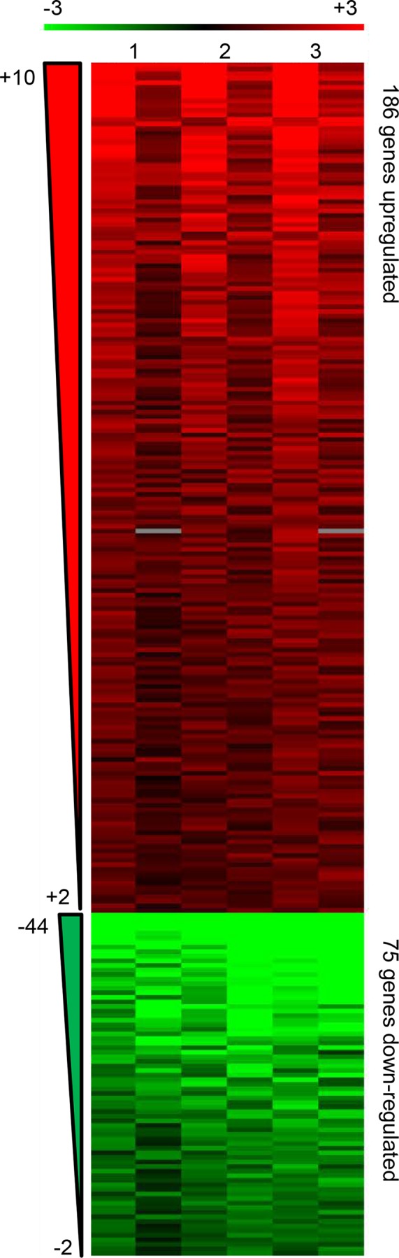
Mycobacterium tuberculosis (M. tb) infection is initiated by the few bacilli inhaled into the alveolus. Studies in lungs of aerosol-infected mice provided evidence for extensive replication of M. tb in non-migrating, non-antigen-presenting cells in the alveoli during the first 2-3 weeks post-infection. Alveoli are lined by type II and type I alveolar epithelial cells (AEC) which outnumber alveolar macrophages by several hundred-fold. M. tb DNA and viable M. tb have been demonstrated in AEC and other non-macrophage cells of the kidney, liver, and spleen in autopsied tissues from latently-infected subjects from TB-endemic regions indicating systemic bacterial dissemination during primary infection. M. tb have also been demonstrated to replicate rapidly in A549 cells (type II AEC line) and acquire increased invasiveness for endothelial cells. Together, these results suggest that AEC could provide an important niche for bacterial expansion and development of a phenotype that promotes dissemination during primary infection. In the current studies, we have compared the transcriptional profile of M. tb replicating intracellularly in A549 cells to that of M. tb replicating in laboratory broth, by microarray analysis. Genes significantly upregulated during intracellular residence were consistent with an active, replicative, metabolic, and aerobic state, as were genes for tryptophan synthesis and for increased virulence (ESAT-6, and ESAT-6-like genes, esxH, esxJ, esxK, esxP, and esxW). In contrast, significant downregulation of the DevR (DosR) regulon and several hypoxia-induced genes was observed. Stress response genes were either not differentially expressed or were downregulated with the exception of the heat shock response and those induced by low pH. The intra-type II AEC M. tb transcriptome strongly suggests that AEC could provide a safe haven in which M. tb can expand dramatically and disseminate from the lung prior to the elicitation of adaptive immune responses.
结核分枝杆菌(M. tb)感染始于少量吸入肺泡的杆菌。对气溶胶感染小鼠肺部的研究表明,在感染后的头2 - 3周内,M. tb在肺泡中不迁移、不呈递抗原的细胞中大量复制。肺泡由II型和I型肺泡上皮细胞(AEC)排列,其数量比肺泡巨噬细胞多出数百倍。在来自结核病流行地区的潜伏感染受试者的尸检组织中,已在肾脏、肝脏和脾脏的AEC及其他非巨噬细胞中检测到M. tb DNA和活的M. tb,这表明在原发性感染期间细菌发生了全身播散。M. tb也已被证明能在A549细胞(II型AEC系)中快速复制,并增强对内皮细胞的侵袭性。这些结果共同表明,AEC可能为细菌扩增以及形成在原发性感染期间促进播散的表型提供重要场所。在当前研究中,我们通过微阵列分析比较了在A549细胞内复制的M. tb与在实验室肉汤中复制的M. tb的转录谱。细胞内生存期间显著上调的基因与活跃、复制、代谢及需氧状态一致,色氨酸合成基因以及毒力增加相关基因(ESAT - 6和ESAT - 6样基因、esxH、esxJ、esxK、esxP和esxW)也是如此。相比之下,观察到DevR(DosR)调控子和几个缺氧诱导基因显著下调。应激反应基因要么没有差异表达,要么除热休克反应及低pH诱导的基因外均下调。II型AEC内的M. tb转录组强烈表明,AEC可为M. tb提供一个安全的避难所,使其能够在适应性免疫反应引发之前在肺部大量扩增并播散。