Hernandez James R, Kim John J, Verdone James E, Liu Xin, Torga Gonzalo, Pienta Kenneth J, Mooney Steven M
Department of Urology, The James Buchanan Brady Urological Institute, Johns Hopkins University, Baltimore, MD, 21287, USA,
Med Oncol. 2015 May;32(5):159. doi: 10.1007/s12032-015-0593-z. Epub 2015 Apr 9.
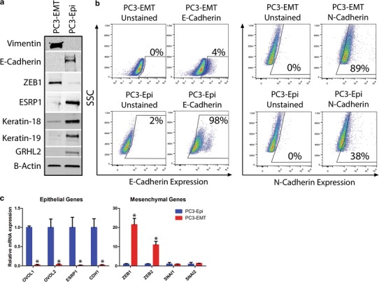
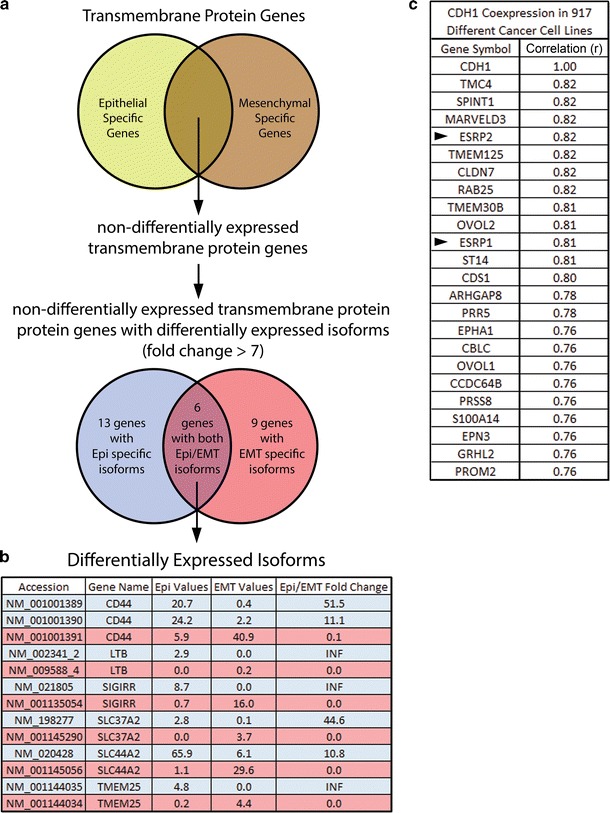
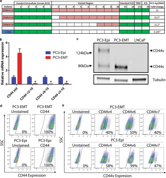
An epithelial to mesenchymal transition (EMT) has been shown to be a necessary precursor to prostate cancer metastasis. Additionally, the differential expression and splicing of mRNAs has been identified as a key means to distinguish epithelial from mesenchymal cells by qPCR, western blotting and immunohistochemistry. However, few markers exist to differentiate between these cells by flow cytometry. We previously developed two cell lines, PC3-Epi (epithelial) and PC3-EMT (mesenchymal). RNAseq was used to determine the differential expression of membrane proteins on PC3-Epi/EMT. We used western blotting, qPCR and flow cytometry to validate the RNAseq results. CD44 was one of six membrane proteins found to be differentially spliced between epithelial and mesenchymal PC3 cells. Although total CD44 was positive in all PC3-Epi/EMT cells, PC3-Epi cells had a higher level of CD44v6 (CD44 variant exon 6). CD44v6 was able to differentiate epithelial from mesenchymal prostate cancer cells using either flow cytometry, western blotting or qPCR.
上皮-间质转化(EMT)已被证明是前列腺癌转移的必要前提。此外,通过定量聚合酶链反应(qPCR)、蛋白质免疫印迹法和免疫组织化学,mRNA的差异表达和剪接已被确定为区分上皮细胞和间质细胞的关键手段。然而,通过流式细胞术区分这些细胞的标志物很少。我们之前开发了两种细胞系,PC3-Epi(上皮细胞)和PC3-EMT(间质细胞)。RNA测序用于确定PC3-Epi/EMT上膜蛋白的差异表达。我们使用蛋白质免疫印迹法、qPCR和流式细胞术来验证RNA测序结果。CD44是在上皮和间质PC3细胞之间发现差异剪接的六种膜蛋白之一。尽管所有PC3-Epi/EMT细胞中的总CD44均为阳性,但PC3-Epi细胞中CD44v6(CD44可变外显子6)的水平更高。CD44v6能够通过流式细胞术、蛋白质免疫印迹法或qPCR区分上皮性和间质性前列腺癌细胞。