• 文献检索
  • 文档翻译
  • 深度研究
  • 学术资讯
  • Suppr Zotero 插件Zotero 插件
  • 邀请有礼
  • 套餐&价格
  • 历史记录
应用&插件
Suppr Zotero 插件Zotero 插件浏览器插件Mac 客户端Windows 客户端微信小程序
定价
高级版会员购买积分包购买API积分包
服务
文献检索文档翻译深度研究API 文档MCP 服务
关于我们
关于 Suppr公司介绍联系我们用户协议隐私条款
关注我们

Suppr 超能文献

核心技术专利:CN118964589B侵权必究
粤ICP备2023148730 号-1Suppr @ 2026

文献检索

告别复杂PubMed语法,用中文像聊天一样搜索,搜遍4000万医学文献。AI智能推荐,让科研检索更轻松。

立即免费搜索

文件翻译

保留排版,准确专业,支持PDF/Word/PPT等文件格式,支持 12+语言互译。

免费翻译文档

深度研究

AI帮你快速写综述,25分钟生成高质量综述,智能提取关键信息,辅助科研写作。

立即免费体验

前交叉韧带重建术后3年的膝关节软骨。对10个膝关节进行的定量T2弛豫测量分析。

Articular cartilage of the knee 3 years after ACL reconstruction. A quantitative T2 relaxometry analysis of 10 knees.

作者信息

Bae Ji-Hoon, Hosseini Ali, Wang Yang, Torriani Martin, Gill Thomas J, Grodzinsky Alan J, Li Guoan

机构信息

a 1 Bioengineering Laboratory, Department of Orthopaedic Surgery , Massachusetts General Hospital and Harvard Medical School, Boston.

e 5 Department of Orthopaedic Surgery, Korea University Guro Hospital , Korea University College of Medicine, Seoul, Republic of Korea.

出版信息

Acta Orthop. 2015;86(5):605-10. doi: 10.3109/17453674.2015.1039426.

DOI:10.3109/17453674.2015.1039426
PMID:25854533
原文链接:https://pmc.ncbi.nlm.nih.gov/articles/PMC4564784/
Abstract

BACKGROUND AND PURPOSE

T1ρ or T2 relaxation imaging has been increasingly used to evaluate the cartilage of the knee. We investigated the cartilage of ACL-reconstructed knees 3 years after surgery using T2 relaxation times.

PATIENTS AND METHODS

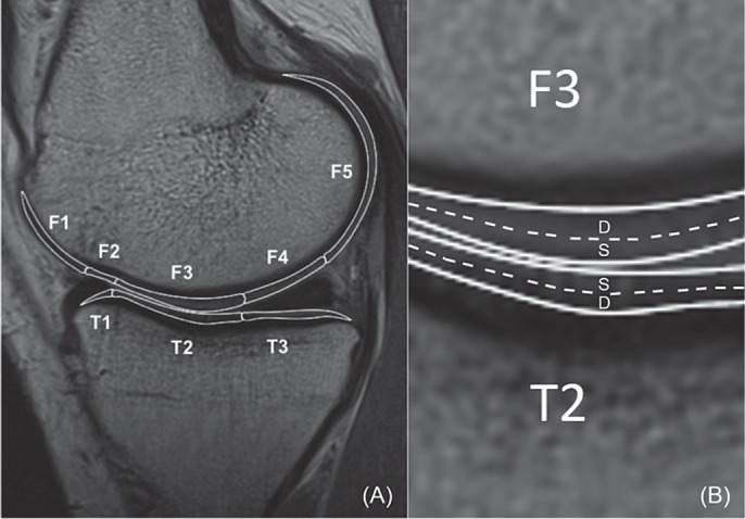
10 patients with a clinically successful unilateral ACL reconstruction were examined 3 years after surgery. Multiple-TE fast-spin echo sagittal images of both knees were acquired using a 3T MRI scanner for T2 mapping of the tibiofemoral cartilage. T2 values of the superficial and deep zones of the tibiofemoral cartilage were analyzed in sub-compartmental areas and compared between the ACL-reconstructed and uninjured contralateral knees.

RESULTS

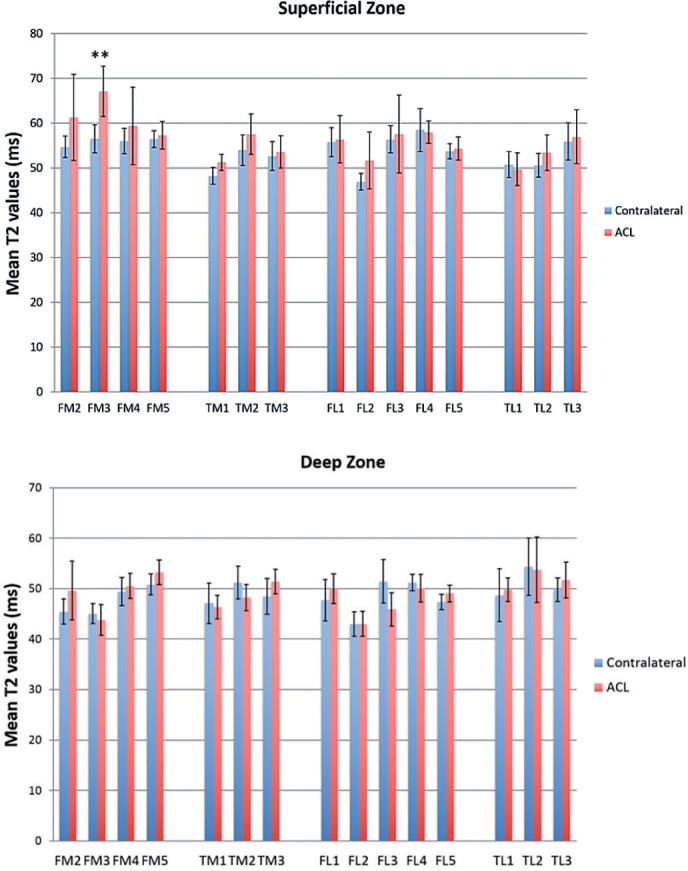
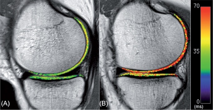
Higher T2 values were observed in 1 or more sub-compartmental areas of each ACL-reconstructed knee compared to the uninjured contralateral side. Most of the T2 increases were observed at the superficial zones of the cartilage, especially at the medial compartment. At the medial compartment of the ACL-reconstructed knee, the T2 values of the femoral and tibial cartilage were increased by 3-81% compared to the uninjured contralateral side, at the superficial zones of the weight-bearing areas. T2 values in the superficial zone of the central medial femoral condyle differed between the 2 groups (p = 0.002).

INTERPRETATION

The articular cartilage of ACL-reconstructed knees, although clinically satisfactory, had higher T2 values in the superficial zone of the central medial femoral condyle than in the uninjured contralateral side 3 years after surgery. Further studies are warranted to determine whether these patients would undergo cartilage degeneration over time.

摘要

背景与目的

T1ρ或T2弛豫成像已越来越多地用于评估膝关节软骨。我们使用T2弛豫时间对前交叉韧带(ACL)重建术后3年的膝关节软骨进行了研究。

患者与方法

对10例临床单侧ACL重建成功的患者在术后3年进行检查。使用3T磁共振成像(MRI)扫描仪获取双膝的多回波快速自旋回波矢状位图像,用于胫股关节软骨的T2成像。分析胫股关节软骨表层和深层区域在亚区域的T2值,并在ACL重建侧与未受伤的对侧膝关节之间进行比较。

结果

与未受伤的对侧相比,每个ACL重建膝关节的1个或更多亚区域观察到更高的T2值。大多数T2值增加出现在软骨的表层区域,尤其是内侧间室。在ACL重建膝关节的内侧间室,与未受伤的对侧相比,在负重区域的表层,股骨和胫骨软骨的T2值增加了3% - 81%。两组之间中央内侧股骨髁表层区域的T2值存在差异(p = 0.002)。

解读

ACL重建膝关节的关节软骨,尽管临床效果良好,但术后3年中央内侧股骨髁表层区域的T2值高于未受伤的对侧。有必要进一步研究以确定这些患者是否会随着时间推移发生软骨退变。

https://cdn.ncbi.nlm.nih.gov/pmc/blobs/5617/4564784/7510cb537805/ORT-86-605-g003.jpg
https://cdn.ncbi.nlm.nih.gov/pmc/blobs/5617/4564784/9610fa788c44/ORT-86-605-g001.jpg
https://cdn.ncbi.nlm.nih.gov/pmc/blobs/5617/4564784/efcc8e379402/ORT-86-605-g002.jpg
https://cdn.ncbi.nlm.nih.gov/pmc/blobs/5617/4564784/7510cb537805/ORT-86-605-g003.jpg
https://cdn.ncbi.nlm.nih.gov/pmc/blobs/5617/4564784/9610fa788c44/ORT-86-605-g001.jpg
https://cdn.ncbi.nlm.nih.gov/pmc/blobs/5617/4564784/efcc8e379402/ORT-86-605-g002.jpg
https://cdn.ncbi.nlm.nih.gov/pmc/blobs/5617/4564784/7510cb537805/ORT-86-605-g003.jpg

相似文献

1
Articular cartilage of the knee 3 years after ACL reconstruction. A quantitative T2 relaxometry analysis of 10 knees.前交叉韧带重建术后3年的膝关节软骨。对10个膝关节进行的定量T2弛豫测量分析。
Acta Orthop. 2015;86(5):605-10. doi: 10.3109/17453674.2015.1039426.
2
Comparison of T1rho relaxation times between ACL-reconstructed knees and contralateral uninjured knees.前交叉韧带重建膝关节与对侧未受伤膝关节之间的T1rho弛豫时间比较。
Knee Surg Sports Traumatol Arthrosc. 2014 Feb;22(2):298-307. doi: 10.1007/s00167-013-2397-z. Epub 2013 Jan 31.
3
Cartilage Subsurface Changes to Magnetic Resonance Imaging UTE-T2* 2 Years After Anterior Cruciate Ligament Reconstruction Correlate With Walking Mechanics Associated With Knee Osteoarthritis.前交叉韧带重建后 2 年的磁共振成像UTE-T2*软骨下变化与与膝关节骨关节炎相关的膝关节行走力学相关。
Am J Sports Med. 2018 Mar;46(3):565-572. doi: 10.1177/0363546517743969. Epub 2018 Jan 2.
4
Cartilage morphology and T1ρ and T2 quantification in ACL-reconstructed knees: a 2-year follow-up.ACL 重建膝关节的软骨形态学及 T1ρ 和 T2 值定量:2 年随访。
Osteoarthritis Cartilage. 2013 Aug;21(8):1058-67. doi: 10.1016/j.joca.2013.05.010. Epub 2013 May 22.
5
Quantitative MRI T2 relaxation time evaluation of knee cartilage: comparison of meniscus-intact and -injured knees after anterior cruciate ligament reconstruction.膝关节软骨的定量MRI T2弛豫时间评估:前交叉韧带重建术后半月板完整与损伤膝关节的比较
Am J Sports Med. 2015 Apr;43(4):865-72. doi: 10.1177/0363546514564151. Epub 2015 Jan 14.
6
Gait Characteristics Associated With a Greater Increase in Medial Knee Cartilage T and T Relaxation Times in Patients Undergoing Anterior Cruciate Ligament Reconstruction.前交叉韧带重建患者中与内侧膝关节软骨T和T弛豫时间更大增加相关的步态特征
Am J Sports Med. 2017 Dec;45(14):3262-3271. doi: 10.1177/0363546517723007. Epub 2017 Sep 12.
7
Quantitative analysis of T2 relaxation times of the patellofemoral joint cartilage 3 years after anterior cruciate ligament reconstruction.前交叉韧带重建术后3年髌股关节软骨T2弛豫时间的定量分析。
J Orthop Translat. 2017 Jul 2;12:85-92. doi: 10.1016/j.jot.2017.06.002. eCollection 2018 Jan.
8
Quantitative evaluation of the tibiofemoral joint cartilage by T2 mapping in patients with acute anterior cruciate ligament injury vs contralateral knees: results from the subacute phase using data from the NACOX study cohort.采用 NACOX 研究队列亚急性期数据的急性前交叉韧带损伤患者与对侧膝关节的 T2 mapping 定量评估:胫骨股骨关节软骨结果。
Osteoarthritis Cartilage. 2022 Jul;30(7):987-997. doi: 10.1016/j.joca.2022.02.623. Epub 2022 Apr 11.
9
Quantitative Magnetic Resonance Imaging UTE-T2* Mapping of Cartilage and Meniscus Healing After Anatomic Anterior Cruciate Ligament Reconstruction.解剖学前交叉韧带重建术后软骨和半月板愈合的定量磁共振成像UTE-T2*映射
Am J Sports Med. 2014 Aug;42(8):1847-56. doi: 10.1177/0363546514532227. Epub 2014 May 8.
10
MRI UTE-T2* shows high incidence of cartilage subsurface matrix changes 2 years after ACL reconstruction.MRI UTE-T2* 显示 ACL 重建 2 年后软骨下基质改变的发生率较高。
J Orthop Res. 2019 Feb;37(2):370-377. doi: 10.1002/jor.24110. Epub 2019 Jan 8.

引用本文的文献

1
Finite Element Models of Osteocytes and Their Load-Induced Activation.成骨细胞及其受力激活的有限元模型。
Curr Osteoporos Rep. 2022 Apr;20(2):127-140. doi: 10.1007/s11914-022-00728-9. Epub 2022 Mar 17.
2
Feasibility of T2 Mapping of the Sacroiliac Joints in Healthy Control Subjects and Children and Young Adults with Sacroiliitis.健康对照受试者以及患有骶髂关节炎的儿童和年轻人中骶髂关节T2映射的可行性
ACR Open Rheumatol. 2022 Jan;4(1):74-82. doi: 10.1002/acr2.11354. Epub 2021 Nov 10.
3
Longitudinal T2 Mapping and Texture Feature Analysis in the Detection and Monitoring of Experimental Post-Traumatic Cartilage Degeneration.

本文引用的文献

1
Early knee osteoarthritis is evident one year following anterior cruciate ligament reconstruction: a magnetic resonance imaging evaluation.前交叉韧带重建术后 1 年即可出现早期膝关节骨关节炎:磁共振成像评估。
Arthritis Rheumatol. 2015 Apr;67(4):946-55. doi: 10.1002/art.39005.
2
Increased risk of osteoarthritis after anterior cruciate ligament reconstruction: a 14-year follow-up study of a randomized controlled trial.前交叉韧带重建后骨关节炎风险增加:一项随机对照试验的 14 年随访研究。
Am J Sports Med. 2014 May;42(5):1049-57. doi: 10.1177/0363546514526139. Epub 2014 Mar 18.
3
Does ACL reconstruction alter natural history?: A systematic literature review of long-term outcomes.
纵向T2映射和纹理特征分析在实验性创伤后软骨退变检测与监测中的应用
Life (Basel). 2021 Mar 5;11(3):201. doi: 10.3390/life11030201.
4
Rapid Knee MRI Acquisition and Analysis Techniques for Imaging Osteoarthritis.用于骨关节炎成像的快速膝关节磁共振成像采集与分析技术
J Magn Reson Imaging. 2020 Nov;52(5):1321-1339. doi: 10.1002/jmri.26991. Epub 2019 Nov 21.
5
Bone Marrow Mesenchymal Stromal Cell Treatment in Patients with Osteoarthritis Results in Overall Improvement in Pain and Symptoms and Reduces Synovial Inflammation.骨髓间充质基质细胞治疗骨关节炎患者可整体改善疼痛和症状,并减轻滑膜炎症。
Stem Cells Transl Med. 2019 Aug;8(8):746-757. doi: 10.1002/sctm.18-0183. Epub 2019 Apr 9.
6
Abnormal Biomechanics at 6 Months Are Associated With Cartilage Degeneration at 3 Years After Anterior Cruciate Ligament Reconstruction.前交叉韧带重建后 6 个月的生物力学异常与 3 年后的软骨退变有关。
Arthroscopy. 2019 Feb;35(2):511-520. doi: 10.1016/j.arthro.2018.07.033. Epub 2018 Nov 22.
7
MRI UTE-T2* shows high incidence of cartilage subsurface matrix changes 2 years after ACL reconstruction.MRI UTE-T2* 显示 ACL 重建 2 年后软骨下基质改变的发生率较高。
J Orthop Res. 2019 Feb;37(2):370-377. doi: 10.1002/jor.24110. Epub 2019 Jan 8.
8
Short-Term Contact Kinematic Changes and Longer-Term Biochemical Changes in the Cartilage After ACL Reconstruction: A Pilot Study.ACL 重建后软骨的短期接触运动学变化和长期生化变化:一项初步研究。
Ann Biomed Eng. 2018 Nov;46(11):1797-1805. doi: 10.1007/s10439-018-2079-6. Epub 2018 Jun 26.
9
Quantitative analysis of T2 relaxation times of the patellofemoral joint cartilage 3 years after anterior cruciate ligament reconstruction.前交叉韧带重建术后3年髌股关节软骨T2弛豫时间的定量分析。
J Orthop Translat. 2017 Jul 2;12:85-92. doi: 10.1016/j.jot.2017.06.002. eCollection 2018 Jan.
10
Quantitative imaging of bone-cartilage interactions in ACL-injured patients with PET-MRI.使用 PET-MRI 对 ACL 损伤患者的骨-软骨相互作用进行定量成像。
Osteoarthritis Cartilage. 2018 Jun;26(6):790-796. doi: 10.1016/j.joca.2018.04.001. Epub 2018 Apr 12.
前交叉韧带重建会改变自然病程吗?:一项关于长期预后的系统文献综述。
J Bone Joint Surg Am. 2014 Feb 19;96(4):292-300. doi: 10.2106/JBJS.L.01713.
4
Quantitative magnetic resonance imaging assessment of cartilage status: a comparison between young men with and without anterior cruciate ligament reconstruction.定量磁共振成像评估软骨状态:前交叉韧带重建的年轻男性与无重建者的比较。
Arthroscopy. 2013 Dec;29(12):2012-9. doi: 10.1016/j.arthro.2013.09.075.
5
Anterior cruciate ligament injury and radiologic progression of knee osteoarthritis: a systematic review and meta-analysis.前交叉韧带损伤与膝关节骨关节炎的影像学进展:一项系统评价和荟萃分析。
Am J Sports Med. 2014 Sep;42(9):2242-52. doi: 10.1177/0363546513508376. Epub 2013 Nov 8.
6
Articular cartilage lesions increase early cartilage degeneration in knees treated by anterior cruciate ligament reconstruction: T1ρ mapping evaluation and 1-year follow-up.关节软骨损伤增加前交叉韧带重建治疗膝关节的早期软骨退变:T1ρ 图谱评估及 1 年随访。
Am J Sports Med. 2013 Oct;41(10):2353-61. doi: 10.1177/0363546513496048. Epub 2013 Aug 7.
7
Cartilage and meniscal T2 relaxation time as non-invasive biomarker for knee osteoarthritis and cartilage repair procedures.软骨和半月板 T2 弛豫时间作为膝关节骨关节炎和软骨修复手术的非侵入性生物标志物。
Osteoarthritis Cartilage. 2013 Oct;21(10):1474-84. doi: 10.1016/j.joca.2013.07.012. Epub 2013 Jul 27.
8
Cartilage morphology and T1ρ and T2 quantification in ACL-reconstructed knees: a 2-year follow-up.ACL 重建膝关节的软骨形态学及 T1ρ 和 T2 值定量:2 年随访。
Osteoarthritis Cartilage. 2013 Aug;21(8):1058-67. doi: 10.1016/j.joca.2013.05.010. Epub 2013 May 22.
9
Cartilage status in relation to return to sports after anterior cruciate ligament reconstruction.前交叉韧带重建术后与重返运动相关的软骨状态。
Am J Sports Med. 2013 Mar;41(3):550-9. doi: 10.1177/0363546512473568. Epub 2013 Feb 4.
10
Comparison of T1rho relaxation times between ACL-reconstructed knees and contralateral uninjured knees.前交叉韧带重建膝关节与对侧未受伤膝关节之间的T1rho弛豫时间比较。
Knee Surg Sports Traumatol Arthrosc. 2014 Feb;22(2):298-307. doi: 10.1007/s00167-013-2397-z. Epub 2013 Jan 31.