González de San Román Estibaliz, Manuel Iván, Giralt María Teresa, Chun Jerold, Estivill-Torrús Guillermo, Rodríguez de Fonseca Fernando, Santín Luis Javier, Ferrer Isidro, Rodríguez-Puertas Rafael
Department of Pharmacology, Faculty of Medicine and Odontology, University of the Basque Country, UPV/EHU, Leioa, Spain.
Molecular and Cellular Neuroscience Department, Dorris Neuroscience Center, The Scripps Research Institute, La Jolla, California, USA.
J Neurochem. 2015 Aug;134(3):471-85. doi: 10.1111/jnc.13112. Epub 2015 Apr 27.
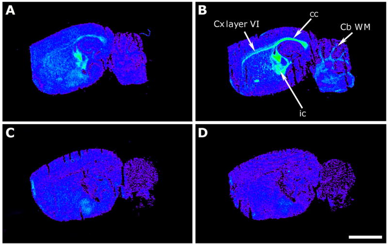
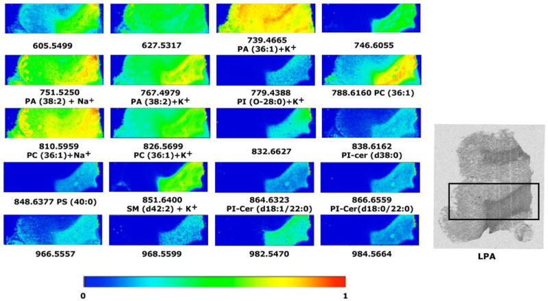
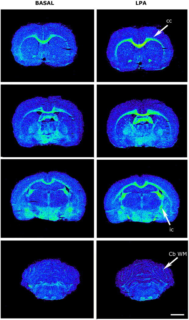
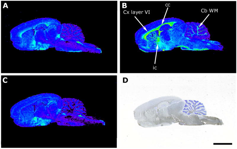
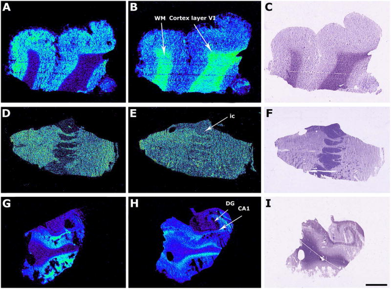
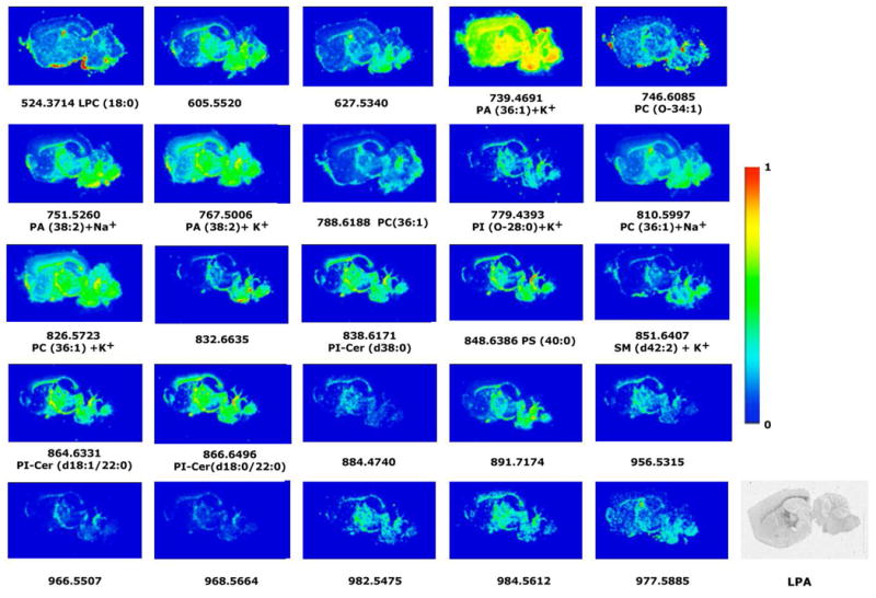
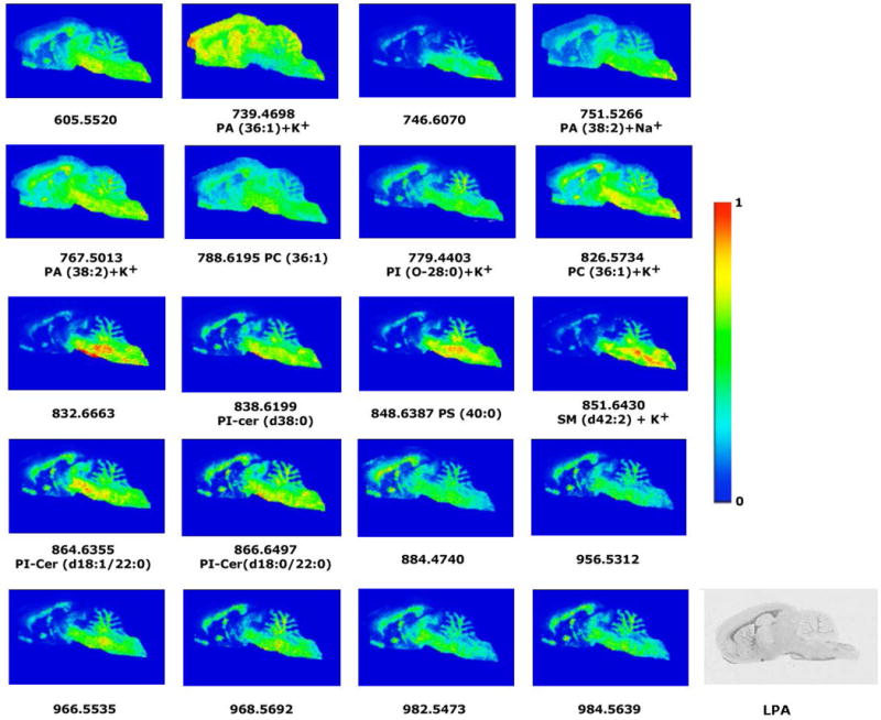
Lysophosphatidic acid (LPA) is a signaling molecule that binds to six known G protein-coupled receptors: LPA1 -LPA6 . LPA evokes several responses in the CNS, including cortical development and folding, growth of the axonal cone and its retraction process. Those cell processes involve survival, migration, adhesion proliferation, differentiation, and myelination. The anatomical localization of LPA1 is incompletely understood, particularly with regard to LPA binding. Therefore, we have used functional [(35) S]GTPγS autoradiography to verify the anatomical distribution of LPA1 binding sites in adult rodent and human brain. The greatest activity was observed in myelinated areas of the white matter such as corpus callosum, internal capsule and cerebellum. MaLPA1 -null mice (a variant of LPA1 -null) lack [(35) S]GTPγS basal binding in white matter areas, where the LPA1 receptor is expressed at high levels, suggesting a relevant role of the activity of this receptor in the most myelinated brain areas. In addition, phospholipid precursors of LPA were localized by MALDI-IMS in both rodent and human brain slices identifying numerous species of phosphatides and phosphatidylcholines. Both phosphatides and phosphatidylcholines species represent potential LPA precursors. The anatomical distribution of these precursors in rodent and human brain may indicate a metabolic relationship between LPA and LPA1 receptors. Lysophosphatidic acid (LPA) is a signaling molecule that binds to six known G protein-coupled receptors (GPCR), LPA1 to LPA6 . LPA evokes several responses in the central nervous system (CNS), including cortical development and folding, growth of the axonal cone and its retraction process. We used functional [(35) S]GTPγS autoradiography to verify the anatomical distribution of LPA1 -binding sites in adult rodent and human brain. The distribution of LPA1 receptors in rat, mouse and human brains show the highest activity in white matter myelinated areas. The basal and LPA-evoked activities are abolished in MaLPA1 -null mice. The phospholipid precursors of LPA are localized by MALDI-IMS. The anatomical distribution of LPA precursors in rodent and human brain suggests a relationship with functional LPA1 receptors.
溶血磷脂酸(LPA)是一种信号分子,可与六种已知的G蛋白偶联受体结合:LPA1-LPA6。LPA在中枢神经系统中引发多种反应,包括皮质发育和折叠、轴突锥体的生长及其回缩过程。这些细胞过程涉及存活、迁移、黏附、增殖、分化和髓鞘形成。LPA1的解剖定位尚未完全明确,尤其是在LPA结合方面。因此,我们使用功能性[(35)S]GTPγS放射自显影技术来验证成年啮齿动物和人类大脑中LPA1结合位点的解剖分布。在白质的髓鞘化区域,如胼胝体、内囊和小脑中观察到最大活性。MaLPA1基因敲除小鼠(LPA1基因敲除的一种变体)在白质区域缺乏[(35)S]GTPγS基础结合,而LPA1受体在该区域高水平表达,这表明该受体的活性在髓鞘化程度最高的脑区中具有重要作用。此外,通过基质辅助激光解吸/电离成像质谱(MALDI-IMS)在啮齿动物和人类脑切片中定位了LPA的磷脂前体,鉴定出了多种磷脂和磷脂酰胆碱。磷脂和磷脂酰胆碱都代表潜在的LPA前体。这些前体在啮齿动物和人类大脑中的解剖分布可能表明LPA与LPA1受体之间存在代谢关系。溶血磷脂酸(LPA)是一种信号分子,可与六种已知的G蛋白偶联受体(GPCR),即LPA1至LPA六结合。LPA在中枢神经系统(CNS)中引发多种反应,包括皮质发育和折叠、轴突锥体的生长及其回缩过程。我们使用功能性[(35)S]GTPγS放射自显影技术来验证成年啮齿动物和人类大脑中LPA1结合位点的解剖分布。LPA1受体在大鼠、小鼠和人类大脑中的分布显示,在白质髓鞘化区域活性最高。在MaLPA1基因敲除小鼠中,基础活性和LPA诱发的活性均被消除。LPA的磷脂前体通过MALDI-IMS进行定位。LPA前体在啮齿动物和人类大脑中的解剖分布表明其与功能性LPA1受体之间存在关联。