Guerra-Pereira InêsI, Vaz Paula, Faria-Almeida Ricardo, Braga Ana-Cristina, Felino António
Department of Oral Surgery, Faculty of Dentistry of Oporto University, Rua Dr. Manuel Pereira da Silva, 4200 - 393 Porto - Portugal,
Med Oral Patol Oral Cir Bucal. 2015 Jul 1;20(4):e419-26. doi: 10.4317/medoral.20513.
Proximity of the dental roots to the sinus floor makes dental disease a probable cause of maxillary sinusitis. The aim of this study was to find out if maxillary sinus pathologic changes were more prevalent in patients with dental disease and to evaluate the performance of computed tomography (CT) in analyzing and detecting apical periodontitis and other odontogenic causes on the maxillary sinusitis etiology in a Portuguese Caucasian population.
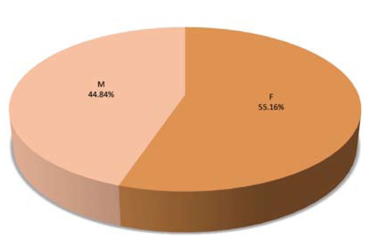
Retrospective cohort study. The total sample of 504 patients and their CT was included in this study. The patients were from a private dental clinic, specializing in oral surgery, where the first complaint was not directly related to sinus disease, but with dental pathology. For each patient, the etiological factors of maxillary sinusitis and the imaging CT findings were analyzed. All the axial, coronal and sagittal CT slices were evaluated and general data were registered. The latter was selected based on the maxillary sinus CT published literature.
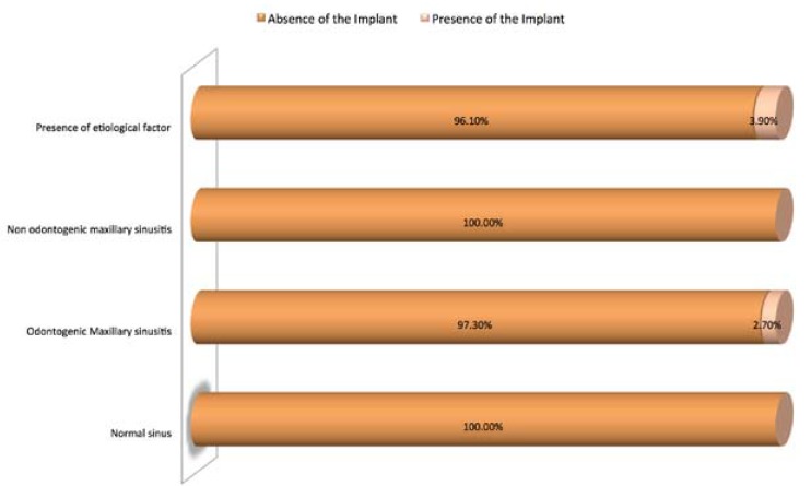
32.40% of patients presented normal sinus (without any etiological factor associated), 29.00% showed presence of etiological and imaging findings in the maxillary sinus, 20.60% had only imaging changes in the maxillary sinus and 18.00% of patients presented only etiological factors and no change in the maxillary sinus.
Radiological imaging is an important tool for establishing the diagnosis of maxillary sinus pathology. These results indicate that the CT scan should be an excellent tool for complement the odontogenic sinusitis diagnosis.
牙根靠近鼻窦底使得牙科疾病可能成为上颌窦炎的病因。本研究的目的是确定上颌窦病理改变在牙科疾病患者中是否更普遍,并评估计算机断层扫描(CT)在分析和检测葡萄牙白种人群上颌窦炎病因中的根尖周炎及其他牙源性病因方面的性能。
回顾性队列研究。本研究纳入了504例患者及其CT数据。患者来自一家专门从事口腔外科的私人牙科诊所,其首要诉求并非直接与鼻窦疾病相关,而是牙科病理问题。对每位患者的上颌窦炎病因及CT影像表现进行分析。评估所有轴向、冠状和矢状位CT切片,并记录一般数据。后者是根据上颌窦CT的已发表文献选取的。
32.40%的患者鼻窦正常(无任何相关病因),29.00%的患者上颌窦存在病因及影像表现,20.60%的患者上颌窦仅有影像改变,18.00%的患者仅有病因因素,上颌窦无变化。
放射影像学是确立上颌窦病理诊断的重要工具。这些结果表明CT扫描应是辅助牙源性鼻窦炎诊断的优秀工具。