Bani-Ata Majid, Aleshawi Abdelwahab, Khatatbeh Abdullah, Al-Domaidat Derar, Alnussair Bayan, Al-Shawaqfeh Raneem, Allouh Mohammed
Otolaryngology Department, Faculty of Medicine, Jordan University of Science and Technology, Irbid 22110, Jordan.
King Abdullah University Hospital, Jordan University of Science and Technology, Irbid 22110, Jordan.
Int J Gen Med. 2020 May 8;13:163-168. doi: 10.2147/IJGM.S253569. eCollection 2020.
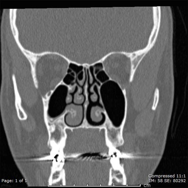
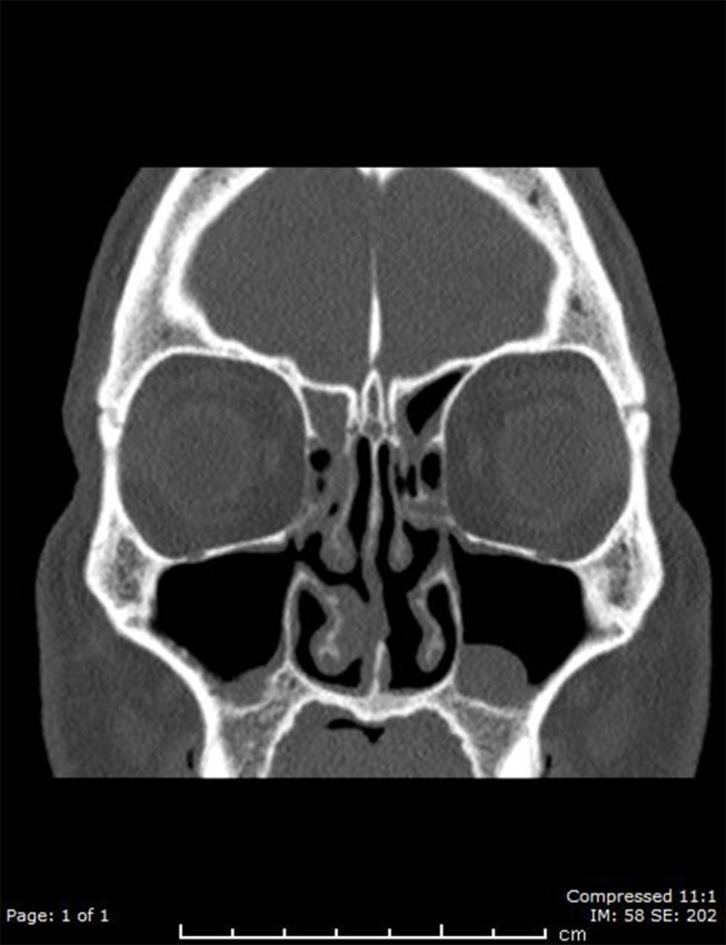
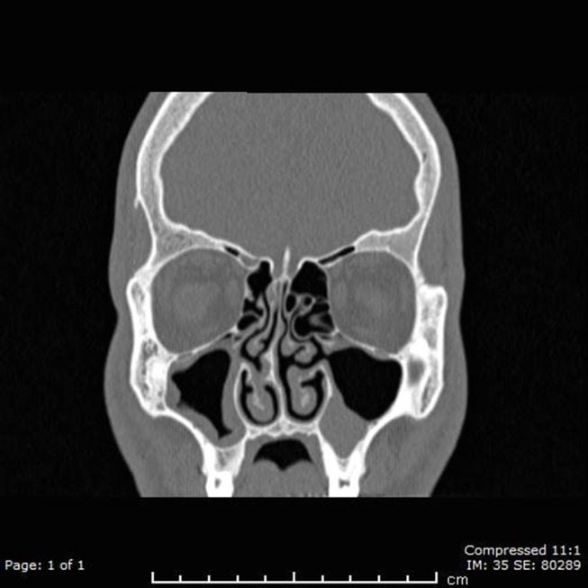
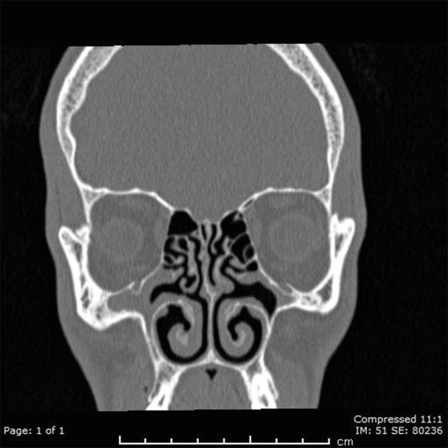
The role of the accessory maxillary ostium, a common anatomical variant, in the development of chronic sinusitis remains unclear. This study aimed to examine the association between chronic sinusitis and presence of an accessory maxillary ostium using computed tomography (CT) of the paranasal sinuses.
We conducted a retrospective study of 1188 paranasal sinus CT scans performed in a major tertiary medical center between January 1, 2016 and December 31, 2016. Axial and coronal and views were reviewed to evaluate the presence of accessory maxillary ostia and maxillary and ethmoid sinusitis.
Nine hundred twenty-eight patients were included for analysis. A 52.8% were male. Mean patient age was 33.8 years. A right accessory maxillary ostium was detected in 274 patients (29.5%), which was the same number of patients with a left accessory maxillary ostium. Bilateral accessory maxillary ostia were found in 172. The presence of right maxillary sinusitis was significantly associated with male gender and the presence of a right accessory maxillary ostium. Male gender was the only factor significantly associated with the presence of left sinusitis. Left or right ethmoidal sinusitis was significantly associated with male gender and the presence of left or right maxillary sinusitis, respectively.
The presence of an accessory maxillary ostium may contribute to the development of maxillary and ethmoidal sinusitis. Further studies are needed to elucidate this association and determine indications for incorporating the natural and accessory ostia when performing middle meatus antrostomy during endoscopic sinus surgery.
副上颌窦口作为一种常见的解剖变异,在慢性鼻窦炎的发生发展中的作用尚不清楚。本研究旨在利用鼻窦计算机断层扫描(CT)检查慢性鼻窦炎与副上颌窦口的存在之间的关联。
我们对2016年1月1日至2016年12月31日在一家大型三级医疗中心进行的1188例鼻窦CT扫描进行了回顾性研究。回顾轴向和冠状位图像以评估副上颌窦口以及上颌窦和筛窦炎的存在情况。
928例患者纳入分析。其中52.8%为男性。患者平均年龄为33.8岁。274例患者(29.5%)检测到右侧副上颌窦口,左侧副上颌窦口的患者数量与之相同。172例患者发现双侧副上颌窦口。右侧上颌窦炎的存在与男性性别以及右侧副上颌窦口的存在显著相关。男性性别是与左侧鼻窦炎存在显著相关的唯一因素。左侧或右侧筛窦炎分别与男性性别以及左侧或右侧上颌窦炎的存在显著相关。
副上颌窦口的存在可能促成上颌窦和筛窦炎的发生。需要进一步研究以阐明这种关联,并确定在内镜鼻窦手术中进行中鼻道上颌窦造口术时纳入自然窦口和副窦口的指征。