Loya-Solis Abelardo, González-Colunga Karla Judith, Pérez-Rodríguez Cynthia M, Ramírez-Ochoa Natalie Sofía, Ceceñas-Falcón Luis, Barboza-Quintana Oralia
Pathology Department, University Hospital "Dr. Jose E. Gonzalez" and Medical School of the Autonomous University of Nuevo Leon, Francisco I. Madero and Gonzalitos, 64460 Monterrey, NL, Mexico.
Otolaryngology-Head and Neck Surgery Department, University Hospital "Dr. Jose E. Gonzalez" and Medical School of the Autonomous University of Nuevo Leon, Francisco I. Madero and Gonzalitos, 64460 Monterrey, NL, Mexico.
Case Rep Pathol. 2015;2015:245026. doi: 10.1155/2015/245026. Epub 2015 Mar 10.
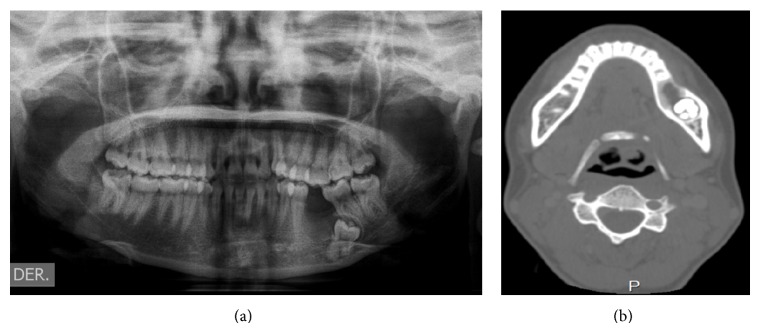
Ameloblastic fibrosarcoma is an uncommon odontogenic tumor composed of a benign epithelial component and a malignant ectomesenchymal component most frequently seen in the third and fourth decades of life. It mainly presents as a painful maxillary or mandibular swelling. Radiographs show a radiolucent mass with ill-defined borders. Radical surgical excision and long-term follow-up are the suggested treatment. We report the case of a 22-year-old female with a 2-month history of an asymptomatic swelling in her left mandible. Examination revealed an exophytic growth measuring 3 × 3 cm extending from the mandibular left first premolar to the second molar. The patient underwent a left hemimandibular resection. Histopathological examination revealed a biphasic tumor composed of inconspicuous islands of benign odontogenic epithelium and an abundant malignant mesenchymal component with marked cellularity, nuclear pleomorphism, hyperchromatism, and moderate mitotic figures with clear margins; one year after the surgical procedure, the patient is clinically and radiologically disease-free.
成釉细胞纤维肉瘤是一种罕见的牙源性肿瘤,由良性上皮成分和恶性外胚间叶成分组成,最常见于30至40岁。它主要表现为上颌或下颌的疼痛性肿胀。X线片显示边界不清的透射性肿块。建议的治疗方法是根治性手术切除和长期随访。我们报告一例22岁女性,左下颌无症状肿胀2个月。检查发现一个从左下颌第一前磨牙延伸至第二磨牙的外生性肿物,大小为3×3厘米。患者接受了左半下颌骨切除术。组织病理学检查显示为双相性肿瘤,由不明显的良性牙源性上皮岛和丰富的恶性间叶成分组成,细胞丰富,有明显的核多形性、核深染,有中度核分裂象且边界清晰;手术后一年,患者临床和影像学检查均无疾病。