Chen Jingyu, Hu Rong, Ge Hongfei, Duanmu Wangsheng, Li Yuhong, Xue Xingseng, Hu Shengli, Feng Hua
Department of Neurosurgery, Southwest Hospital, Third Military Medical University, Chongqing 400038, P.R. China.
Mol Med Rep. 2015 Aug;12(2):1733-40. doi: 10.3892/mmr.2015.3601. Epub 2015 Apr 8.
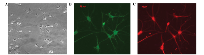
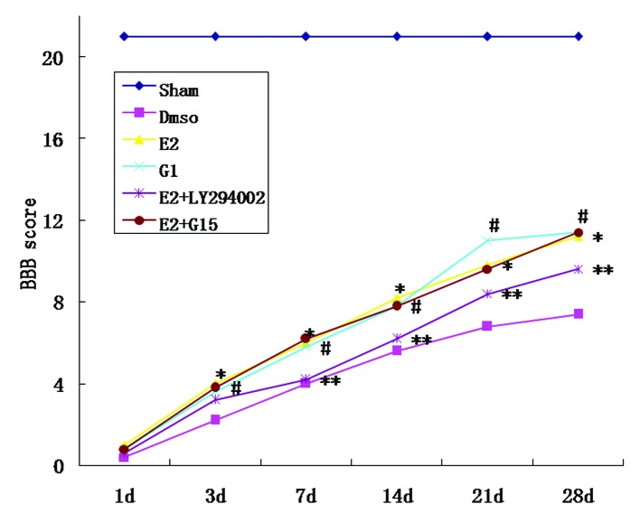
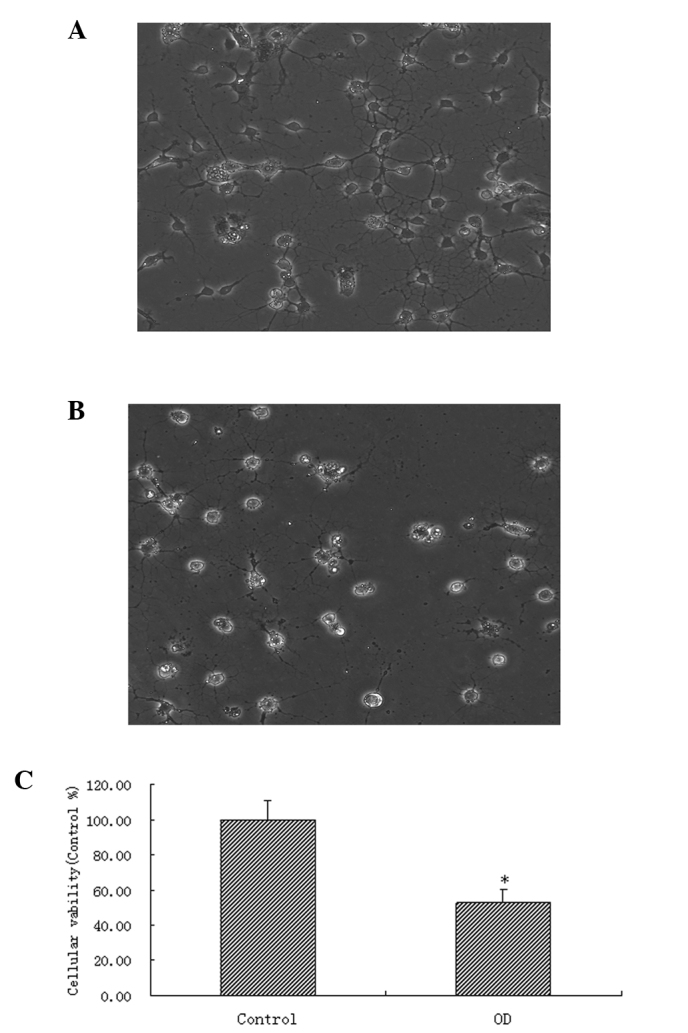
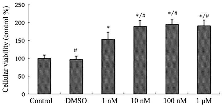
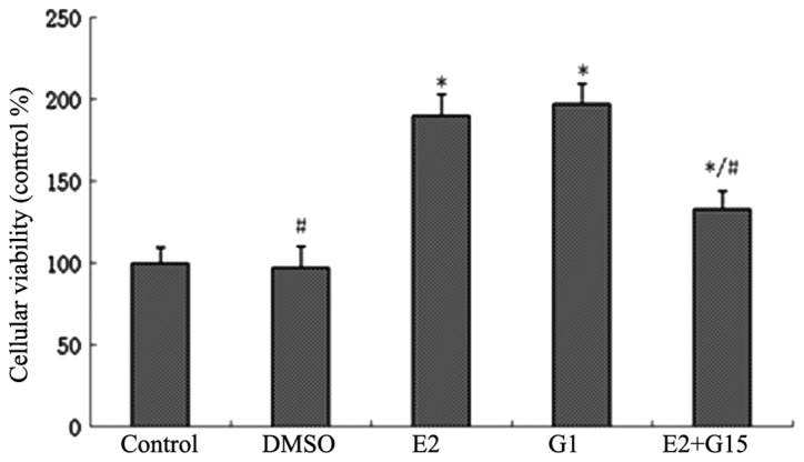
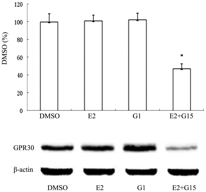
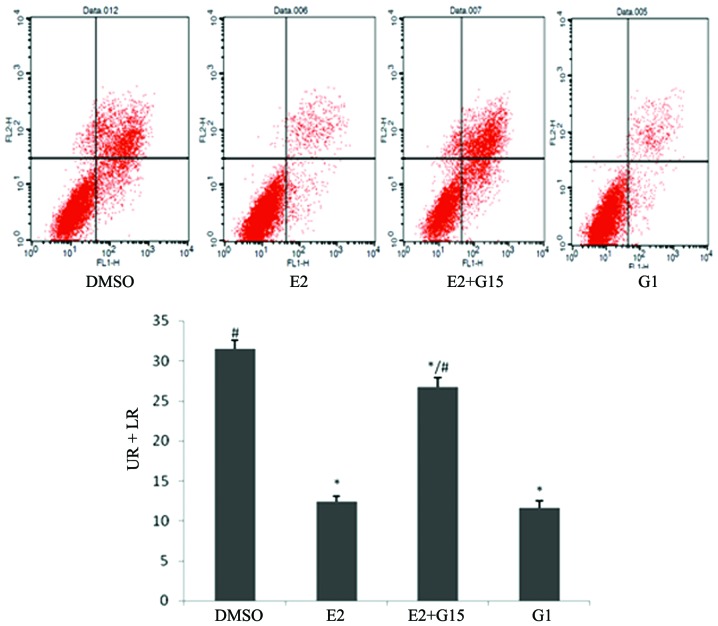
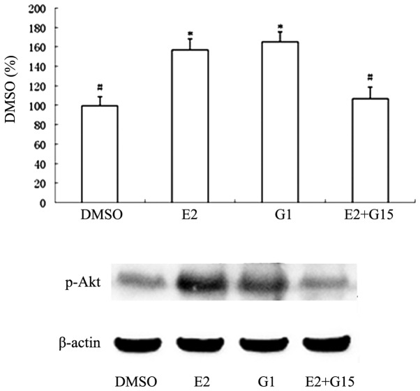
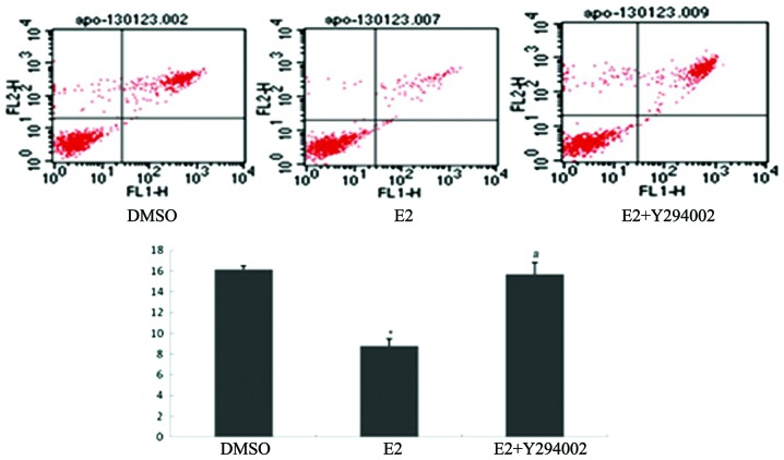
Spinal cord injury (SCI) may result in severe dysfunction of motor neurons. G-protein-coupled receptor 30 (GPR30) expression in the motor neurons of the ventral horn of the spinal cord mediates neuroprotection through estrogen signaling. The present study explored the antiapoptotic effect of estrogen, mediated by GPR30 following SCI, and the mechanisms underlying this effect. Spinal motor neurons from rats were cultured in vitro in order to establish cell models of oxygen and glucose deprivation (OGD). The effects of estrogen, the estrogen agonist, G1, and the estrogen inhibitor, G15, on motor neurons were observed using MTT assays. The effects of E2, G1 and G15 on spinal motor neuron apoptosis following OGD, were detected using flow cytometry. The role of the phosphatidylinositol 3-kinase/protein kinase B (PI3K/Akt) inhibitor, LY294002, was also determined using flow cytometry. Rat SCI models were established. E2, G1 and E2+LY294002 were administered in vivo. Motor function was scored at 3, 7, 14, 21 and 28 d following injury, using Basso-Beattie-Bresnahan (BBB) standards. Cell activity in the estrogen and G1 groups was higher than that in the solvent group, whereas cell activity in the E2+G15 group was lower than that in the E2 group (P<0.05). Following OGD, the proportion of apoptotic cells significantly increased (P<0.05). The proportion in the estrogen group was significantly lower than that in the solvent group, whereas the proportion of apoptotic cells in the E2+G15 and E2+LY294002 groups was higher than that in the E2 group (P<0.05). Treatment with E2 and G1 led to upregulation of P-Akt expression in normal cells and post-OGD cells. The BBB scores of rats in the E2 and G1 groups were higher than those in the placebo group (P<0.05). The BBB scores of the E2+LY294002 group were lower than those of the E2 group (P<0.05). Estrogen thus appears to exert a protective effect on spinal motor neurons following OGD, via GPR30. The PI3K/Akt pathway may be one of those involved in the estrogen‑related antiapoptotic effects mediated by GPR30.
脊髓损伤(SCI)可能导致运动神经元严重功能障碍。脊髓腹角运动神经元中G蛋白偶联受体30(GPR30)的表达通过雌激素信号介导神经保护作用。本研究探讨了SCI后由GPR30介导的雌激素的抗凋亡作用及其作用机制。体外培养大鼠脊髓运动神经元以建立氧糖剥夺(OGD)细胞模型。使用MTT法观察雌激素、雌激素激动剂G1和雌激素抑制剂G15对运动神经元的影响。使用流式细胞术检测E2、G1和G15对OGD后脊髓运动神经元凋亡的影响。还使用流式细胞术确定磷脂酰肌醇3激酶/蛋白激酶B(PI3K/Akt)抑制剂LY294002的作用。建立大鼠SCI模型。在体内给予E2、G1和E2 + LY294002。损伤后3、7、14、21和28天,使用Basso-Beattie-Bresnahan(BBB)标准对运动功能进行评分。雌激素组和G1组的细胞活性高于溶剂组,而E2 + G15组的细胞活性低于E2组(P<0.05)。OGD后,凋亡细胞比例显著增加(P<0.05)。雌激素组的比例显著低于溶剂组,而E2 + G15组和E2 + LY294002组的凋亡细胞比例高于E2组(P<0.05)。E2和G1处理导致正常细胞和OGD后细胞中P-Akt表达上调。E2组和G1组大鼠的BBB评分高于安慰剂组(P<0.05)。E2 + LY294002组的BBB评分低于E2组(P<0.05)。因此,雌激素似乎通过GPR30对OGD后的脊髓运动神经元发挥保护作用。PI3K/Akt途径可能是参与GPR30介导的雌激素相关抗凋亡作用的途径之一。