Chin Man Pan, Chu Patrick H W, Cheong Allen M Y, Chan Henry H L
Laboratory of Experimental Optometry (Neuroscience), School of Optometry, Hong Kong Polytechnic University, Hong Kong SAR, China.
PLoS One. 2015 Apr 13;10(4):e0123480. doi: 10.1371/journal.pone.0123480. eCollection 2015.
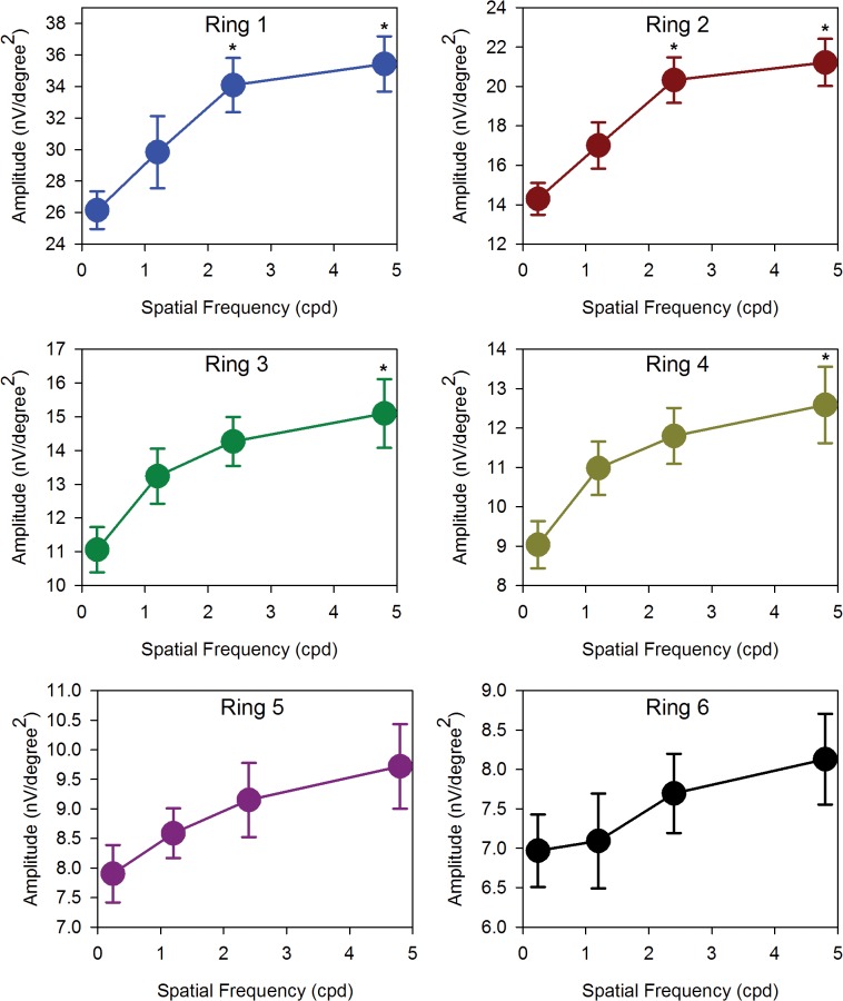
The electrical response of the retina was examined as a function of retinal region, using stimuli of various spatial frequencies in the first experiment. In the second experiment, the regional response of the retina to defocus at high and low spatial frequencies was investigated. Twenty three subjects were recruited for global flash multifocal electroretinogram (mfERG) in experiment 1. Black and white gratings (printed on plastic transparent sheets) of four spatial frequencies (SF), 0.24, 1.2, 2.4 and 4.8 cycle per degree were presented in front of the mfERG stimulation. The amplitudes and implicit times of the direct (DC) and induced (IC) components of mfERG responses were pooled into six concentric rings for analysis. There was low amplitude DC at low SF, which increased with increasing SF, and which decreased with increasing eccentricity. The IC was high in amplitude at all SF and reduced in amplitude with increasing eccentricity. Our findings suggested that outer and inner retina had different characteristics in processing spatial details. In experiment 2, Twenty-three young adults were recruited for mfERG measurement. The retinal electrical responses for low (0.24cpd) and high (4.8cpd) SF under fully corrected conditions of short-term negative defocus (-2D) and short term positive defocus (+2D) conditions were measured. There was a sign-dependent response to defocus in the DC response, mainly in peripheral regions. The sign dependent response at low SF was more obvious than that at high SF, and was located more peripherally. The IC response showed no clear trends for either defocus condition. The human retina seems to have a decoding system for optical defocus, which was tuned for low spatial frequency, and was located in the retinal near periphery.
在第一个实验中,使用各种空间频率的刺激,研究视网膜的电反应与视网膜区域的关系。在第二个实验中,研究了视网膜在高空间频率和低空间频率下对散焦的区域反应。在实验1中,招募了23名受试者进行全视野闪光多焦视网膜电图(mfERG)检查。在mfERG刺激前呈现了四种空间频率(SF)为0.24、1.2、2.4和4.8周/度的黑白光栅(印在塑料透明片上)。将mfERG反应的直接(DC)和诱导(IC)成分的振幅和隐含时间汇总到六个同心环中进行分析。在低空间频率下,DC振幅较低,随空间频率增加而增加,随离心率增加而降低。在所有空间频率下,IC振幅较高,且随离心率增加而降低。我们的研究结果表明,视网膜外层和内层在处理空间细节方面具有不同的特征。在实验2中,招募了23名年轻成年人进行mfERG测量。测量了在短期负性散焦(-2D)和短期正性散焦(+2D)的完全矫正条件下,低(0.24周/度)和高(4.8周/度)空间频率下的视网膜电反应。DC反应中存在散焦的符号依赖性反应,主要在周边区域。低空间频率下的符号依赖性反应比高空间频率下更明显,且位于更周边的位置。IC反应在两种散焦条件下均未显示出明显趋势。人类视网膜似乎有一个用于光学散焦的解码系统,该系统针对低空间频率进行了调整,且位于视网膜近周边区域。