Dipartimento di Scienze Cardiovascolari, Respiratorie, Nefrologiche, Anestesiologiche e Geriatriche, Sapienza-Università di Roma, Rome, Italy; Centro per le Cardiomiopatie Ospedale S. Camillo-Forlanini, Roma, Italy.
Dipartimento di Scienze Cardiovascolari, Respiratorie, Nefrologiche, Anestesiologiche e Geriatriche, Sapienza-Università di Roma, Rome, Italy; Dipartimento di Scienze, Università Roma Tre, Rome, Italy; Center for Evolutionary Ecology, Rome, Italy; Dipartimento di Ingegneria Strutturale e Geotecnica, Sapienza-Università di Roma, Rome, Italy.
PLoS One. 2015 Apr 13;10(4):e0122376. doi: 10.1371/journal.pone.0122376. eCollection 2015.
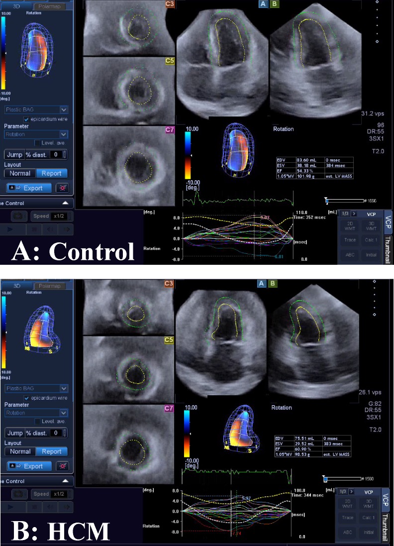
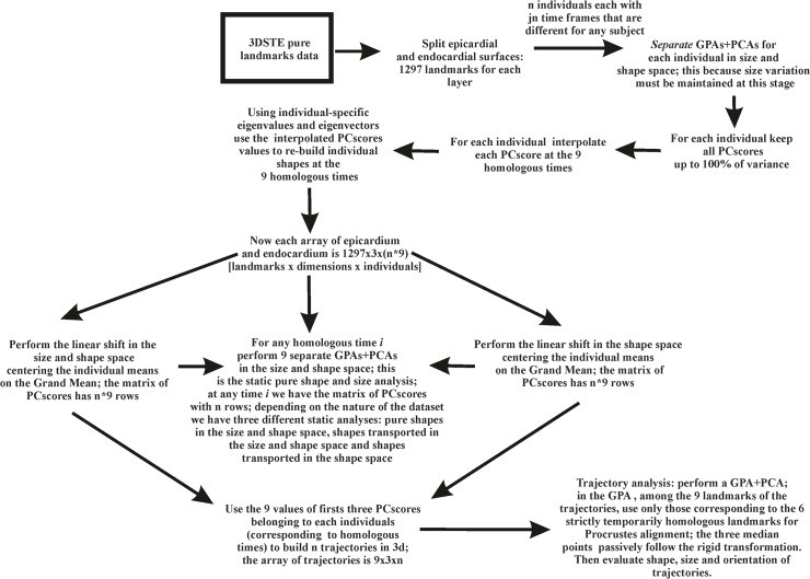
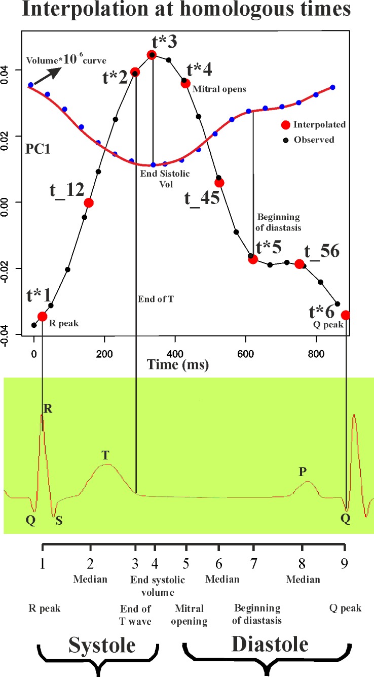
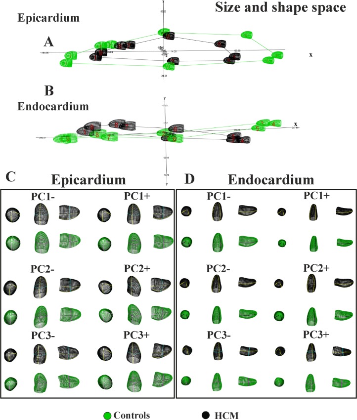
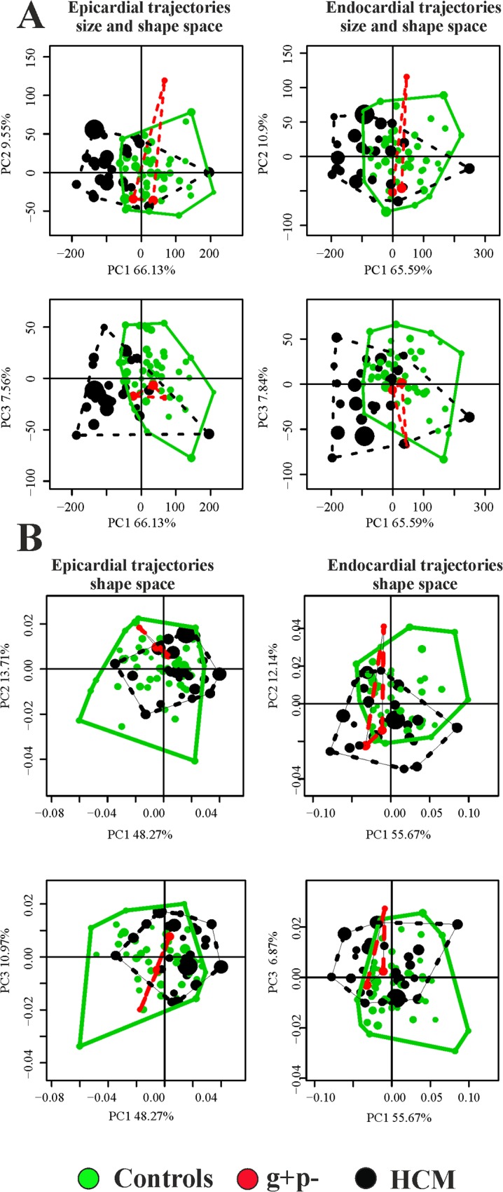
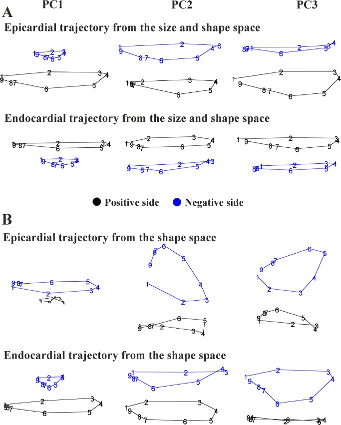
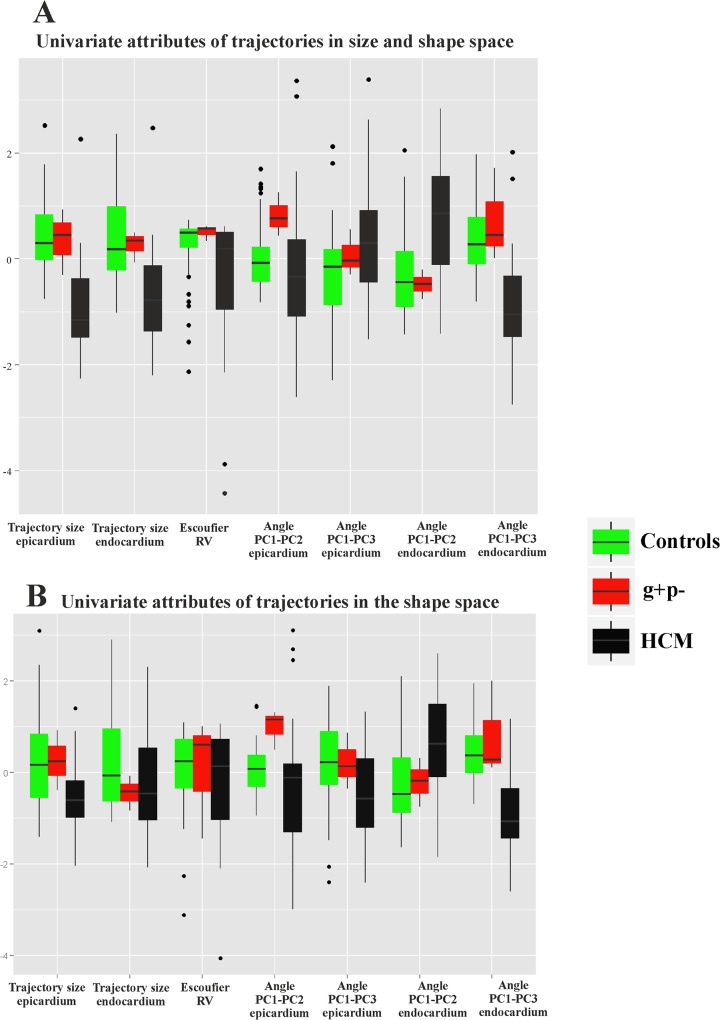
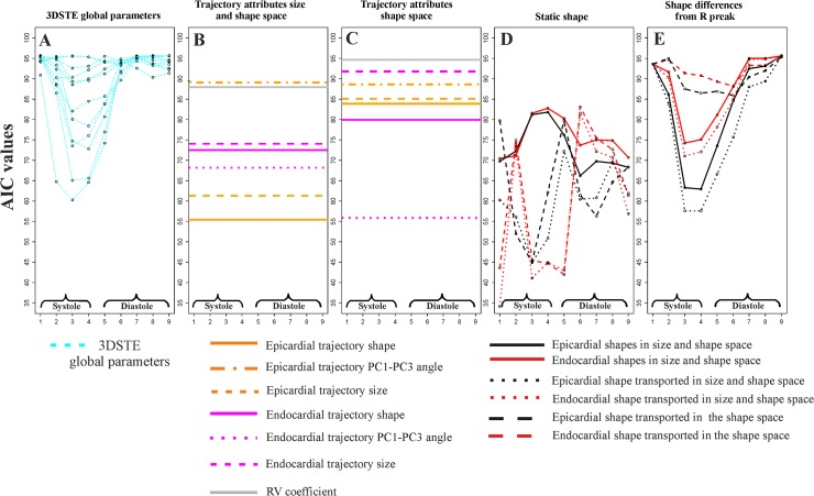
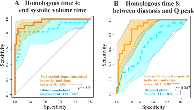
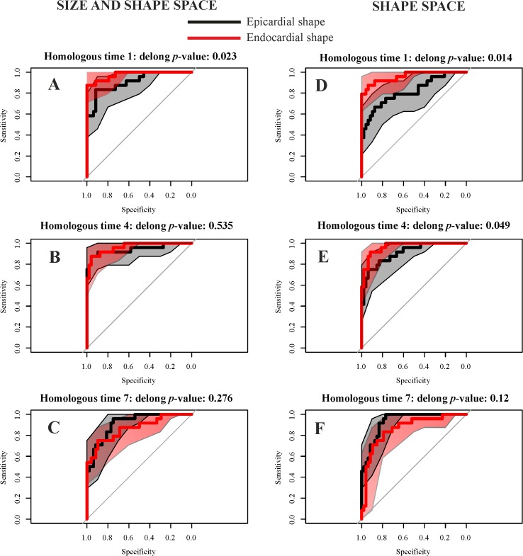
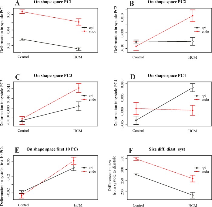
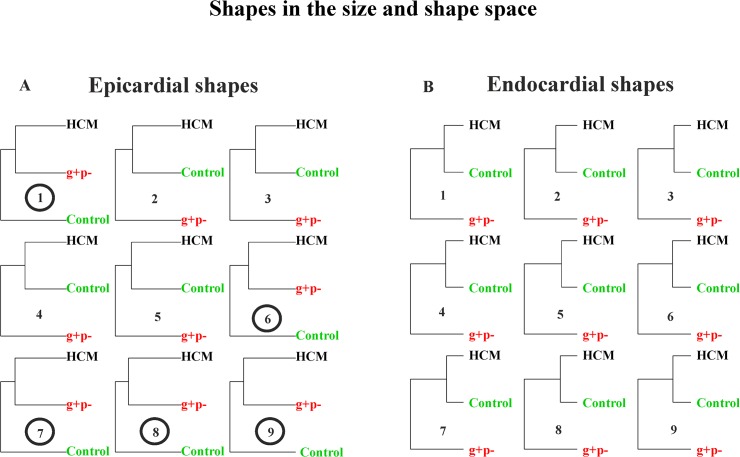
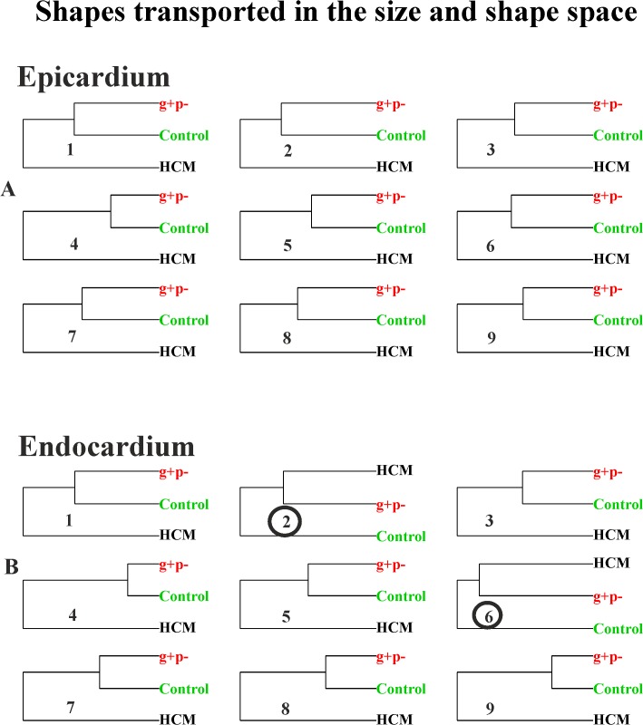
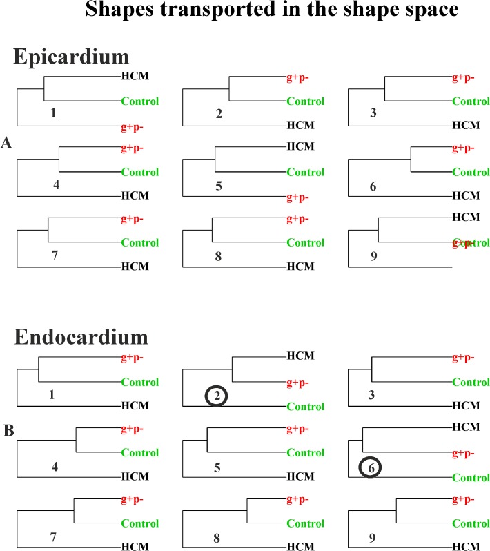
The assessment of left ventricular shape changes during cardiac revolution may be a new step in clinical cardiology to ease early diagnosis and treatment. To quantify these changes, only point registration was adopted and neither Generalized Procrustes Analysis nor Principal Component Analysis were applied as we did previously to study a group of healthy subjects. Here, we extend to patients affected by hypertrophic cardiomyopathy the original approach and preliminarily include genotype positive/phenotype negative individuals to explore the potential that incumbent pathology might also be detected. Using 3D Speckle Tracking Echocardiography, we recorded left ventricular shape of 48 healthy subjects, 24 patients affected by hypertrophic cardiomyopathy and 3 genotype positive/phenotype negative individuals. We then applied Generalized Procrustes Analysis and Principal Component Analysis and inter-individual differences were cleaned by Parallel Transport performed on the tangent space, along the horizontal geodesic, between the per-subject consensuses and the grand mean. Endocardial and epicardial layers were evaluated separately, different from many ecocardiographic applications. Under a common Principal Component Analysis, we then evaluated left ventricle morphological changes (at both layers) explained by first Principal Component scores. Trajectories' shape and orientation were investigated and contrasted. Logistic regression and Receiver Operating Characteristic curves were used to compare these morphometric indicators with traditional 3D Speckle Tracking Echocardiography global parameters. Geometric morphometrics indicators performed better than 3D Speckle Tracking Echocardiography global parameters in recognizing pathology both in systole and diastole. Genotype positive/phenotype negative individuals clustered with patients affected by hypertrophic cardiomyopathy during diastole, suggesting that incumbent pathology may indeed be foreseen by these methods. Left ventricle deformation in patients affected by hypertrophic cardiomyopathy compared to healthy subjects may be assessed by modern shape analysis better than by traditional 3D Speckle Tracking Echocardiography global parameters. Hypertrophic cardiomyopathy pathophysiology was unveiled in a new manner whereby also diastolic phase abnormalities are evident which is more difficult to investigate by traditional ecocardiographic techniques.
评估心脏旋转过程中左心室形状的变化可能是临床心脏病学的一个新步骤,可以方便早期诊断和治疗。为了量化这些变化,我们只采用了点配准,而没有应用广义 Procrustes 分析或主成分分析,就像我们之前研究一组健康受试者时那样。在这里,我们将原始方法扩展到肥厚型心肌病患者,并初步纳入基因型阳性/表型阴性个体,以探索潜在的病理变化也可能被检测到的可能性。使用 3D 斑点追踪超声心动图,我们记录了 48 名健康受试者、24 名肥厚型心肌病患者和 3 名基因型阳性/表型阴性个体的左心室形状。然后,我们应用了广义 Procrustes 分析和主成分分析,并通过在主体共识与总体均值之间的水平测地线的切空间上执行平行传输,清除了个体间的差异。我们分别评估了心内膜和心外膜层,这与许多超声心动图应用不同。在共同的主成分分析下,我们评估了由第一主成分得分解释的左心室形态变化(在心内膜和心外膜层)。研究并对比了轨迹的形状和方向。逻辑回归和接收器工作特征曲线用于比较这些形态计量学指标与传统的 3D 斑点追踪超声心动图全局参数。几何形态计量学指标在识别收缩期和舒张期的病理变化方面优于 3D 斑点追踪超声心动图全局参数。在舒张期,基因型阳性/表型阴性个体与肥厚型心肌病患者聚类,这表明这些方法确实可以预见现有病理。与传统的 3D 斑点追踪超声心动图全局参数相比,肥厚型心肌病患者的左心室变形可以通过现代形状分析更好地评估。肥厚型心肌病的病理生理学以一种新的方式被揭示,其中舒张期异常也很明显,这比传统的超声心动图技术更难研究。