Röling Maarten A, Visser Monique I, Oei Edwin H G, Pilot Peter, Kleinrensink Gert-Jan, Bloem Rolf M
Department of Orthopedic surgery, Reinier de Graafweg 3-11, 2526 AD, Delft, Netherlands.
Department of Radiology Erasmus Medical Center, Rotterdam, Netherlands.
BMC Musculoskelet Disord. 2015 Mar 11;16:50. doi: 10.1186/s12891-015-0504-7.
Femoroacetabular impingement (FAI) is caused by an anatomic deviation of the acetabular rim or proximal femur, which causes chronic groin pain. Radiological identification of FAI can be challenging. Advances in imaging techniques with the use of computed tomography (CT) scan enable 3D simulation of FAI. We made an experimental cadaveric validation study to validate the 3D simulation imaging software.
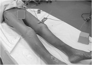
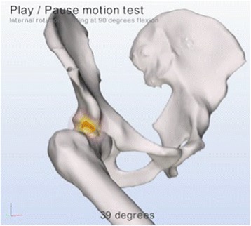
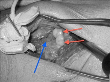
The range of motion (ROM) of five cadaveric hips was measured using an electromagnetic tracking system (EMTS). Specific marked spots in the femur and pelvis were created as reproducible EMTS registration points. Reproducible motions were measured. Hips were subsequently imaged using high-resolution CT after introduction of artificial cam deformities. A proprietary software tool was used, Articulis (Clinical Graphics) to simulate the ROM during the presence and absence of the induced cam deformities.
According to the EMTS, 13 of the 30 measured ROM end-points were restricted by > 5° due to the induced cam deformities. Using Articulis, with the same 5° threshold, we correctly detected 12 of these 13 end point limitations and detected no false positives. The median error of the measured limitations was 1.9° (interquartile range 1.1° - 4.4°). The maximum absolute error was 5.4°.
The use of this dynamic simulation software to determine the presence of motion limiting deformities of the femoroacetabular is validated. The simulation software is able to non-invasively detect a reduction in achievable ROM, caused by a cam type deformity.
股骨髋臼撞击症(FAI)由髋臼边缘或股骨近端的解剖学偏差引起,可导致慢性腹股沟疼痛。FAI的影像学识别可能具有挑战性。计算机断层扫描(CT)扫描等成像技术的进步使得能够对FAI进行三维模拟。我们进行了一项实验性尸体验证研究,以验证三维模拟成像软件。
使用电磁跟踪系统(EMTS)测量五具尸体髋关节的活动范围(ROM)。在股骨和骨盆上创建特定的标记点作为可重复的EMTS注册点。测量可重复的运动。随后在引入人工凸轮畸形后,使用高分辨率CT对髋关节进行成像。使用一种专有软件工具Articulis(Clinical Graphics)来模拟在存在和不存在诱导凸轮畸形时的ROM。
根据EMTS,在30个测量的ROM端点中,有13个由于诱导凸轮畸形而受限超过5°。使用Articulis,在相同的5°阈值下,我们正确检测到了这13个端点限制中的12个,且未检测到假阳性。测量限制的中位数误差为1.9°(四分位间距为1.1° - 4.4°)。最大绝对误差为5.4°。
使用这种动态模拟软件来确定股骨髋臼运动受限畸形的存在得到了验证。该模拟软件能够非侵入性地检测出由凸轮型畸形导致的可实现ROM的降低。