Pishko Gregory L, Muldoon Leslie L, Pagel Michael A, Schwartz Daniel L, Neuwelt Edward A
Fluids Barriers CNS. 2015 Feb 17;12:5. doi: 10.1186/2045-8118-12-5.
Blockade of vascular endothelial growth factor (VEGF) to promote vascular normalization and inhibit angiogenesis has been proposed for the treatment of brain metastases; however, vascular normalization has not been well-characterized in this disease. We investigated the effect of treatment with bevacizumab anti-VEGF antibody on magnetic resonance imaging (MRI) biomarkers of brain tumor vascular characteristics in comparison to small molecule delivery in a rat model of human lung cancer brain metastasis.
Athymic rats with A549 human lung adenocarcinoma intracerebral xenografts underwent MRI at 11.75 T before and one day after treatment with bevacizumab (n = 8) or saline control (n = 8) to evaluate tumor volume, free water content (edema), blood volume and vascular permeability (Ktrans). One day later, permeability to 14C-aminoisobutyric acid (AIB) was measured in tumor and brain to assess the penetration of a small drug-like molecule.
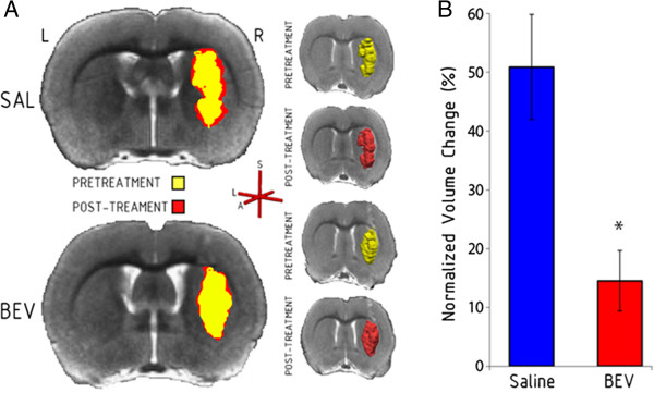
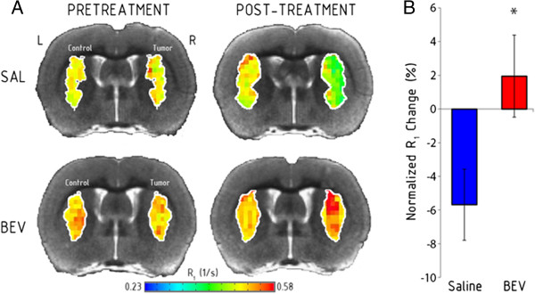
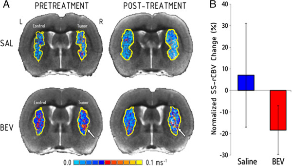
In saline control animals, tumor volume, edema and permeability increased over the two day assessment period. Compared to controls, bevacizumab treatment slowed the rate of tumor growth (P = 0.003) and blocked the increase in edema (P = 0.033), but did not alter tumor blood volume. Bevacizumab also significantly reduced Ktrans (P = 0.033) and AIB passive permeability in tumor (P = 0.04), but not to peritumoral tissue or normal brain. Post-treatment Ktrans correlated with AIB levels in the bevacizumab-treated rats but not in the saline controls.
The correlation of an MRI biomarker for decreased vascular permeability with decreased AIB concentration in tumor after antiangiogenic treatment suggests that bevacizumab partially restored the normal low permeability characteristics of the blood-brain barrier in a model of human lung cancer brain metastasis.
血管内皮生长因子(VEGF)阻断疗法可促进血管正常化并抑制血管生成,已被提议用于治疗脑转移瘤;然而,在这种疾病中血管正常化尚未得到充分描述。我们在人肺癌脑转移大鼠模型中,比较了贝伐单抗抗VEGF抗体治疗与小分子递送对脑肿瘤血管特征磁共振成像(MRI)生物标志物的影响。
将接种A549人肺腺癌的无胸腺大鼠在11.75T磁场下,于接受贝伐单抗治疗(n = 8)或生理盐水对照(n = 8)前及治疗后一天进行MRI检查,以评估肿瘤体积、自由水含量(水肿)、血容量和血管通透性(Ktrans)。一天后,测量肿瘤和脑组织对14C-氨基异丁酸(AIB)的通透性,以评估一种小分子类药物分子的渗透情况。
在生理盐水对照动物中,在两天的评估期内肿瘤体积、水肿和通透性均增加。与对照组相比,贝伐单抗治疗减缓了肿瘤生长速度(P = 0.003)并阻止了水肿增加(P = 0.033),但未改变肿瘤血容量。贝伐单抗还显著降低了肿瘤的Ktrans(P = 0.033)和AIB被动通透性(P = 0.04),但对瘤周组织或正常脑组织无此作用。治疗后的Ktrans与贝伐单抗治疗大鼠的AIB水平相关,但与生理盐水对照组无关。
抗血管生成治疗后,MRI生物标志物显示血管通透性降低与肿瘤中AIB浓度降低相关,这表明在人肺癌脑转移模型中,贝伐单抗部分恢复了血脑屏障正常的低通透性特征。