Fu Cynthia H Y, Costafreda Sergi G, Sankar Anjali, Adams Tracey M, Rasenick Mark M, Liu Peng, Donati Robert, Maglanoc Luigi A, Horton Paul, Marangell Lauren B
School of Psychology, University of East London, Arthur Edwards Building, Rm 3.11, Water Lane, London, E15 4LZ, UK.
Centre for Affective Disorders, Institute of Psychiatry, Psychology and Neuroscience (IoPPN), King's College London, London, UK.
BMC Psychiatry. 2015 Apr 14;15:82. doi: 10.1186/s12888-015-0457-2.
Longitudinal neuroimaging studies of major depressive disorder (MDD) have most commonly assessed the effects of antidepressants from the serotonin reuptake inhibitor class and usually reporting a single measure. Multimodal neuroimaging assessments were acquired from MDD patients during an acute depressive episode with serial measures during a 12-week treatment with the serotonin-norepinephrine reuptake inhibitor (SNRI) duloxetine.
Participants were medication-free MDD patients (n = 32; mean age 40.2 years) in an acute depressive episode and healthy controls matched for age, gender, and IQ (n = 25; mean age 38.8 years). MDD patients received treatment with duloxetine 60 mg daily for 12 weeks with an optional dose increase to 120 mg daily after 8 weeks. All participants had serial imaging at weeks 0, 1, 8, and 12 on a 3 Tesla magnetic resonance imaging (MRI) scanner. Neuroimaging tasks included emotional facial processing, negative attentional bias (emotional Stroop), resting state functional MRI and structural MRI.
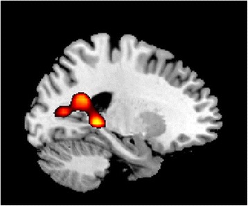
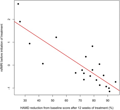
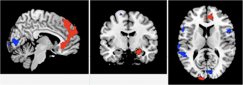
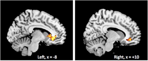
A significant group by time interaction was identified in the anterior default mode network in which MDD patients showed increased connectivity with treatment, while there were no significant changes in healthy participants. In the emotional Stroop task, increased posterior cingulate activation in MDD patients normalized following treatment. No significant group by time effects were observed for happy or sad facial processing, including in amygdala responsiveness, or in regional cerebral volumes. Reduced baseline resting state connectivity within the orbitofrontal component of the default mode network was predictive of clinical response. An early increase in hippocampal volume was predictive of clinical response.
Baseline resting state functional connectivity was predictive of subsequent clinical response. Complementary effects of treatment were observed from the functional neuroimaging correlates of affective facial expressions, negative attentional bias, and resting state. No significant effects were observed in affective facial processing, while the interaction effect in negative attentional bias and individual group effects in resting state connectivity could be related to the SNRI class of antidepressant medication. The specificity of the observed effects to SNRI pharmacological treatments requires further investigation.
Registered at clinicaltrials.gov ( NCT01051466 ).
重度抑郁症(MDD)的纵向神经影像学研究最常评估血清素再摄取抑制剂类抗抑郁药的效果,且通常报告单一测量结果。在重度抑郁症患者的急性抑郁发作期间进行了多模态神经影像学评估,并在使用血清素-去甲肾上腺素再摄取抑制剂(SNRI)度洛西汀进行12周治疗期间进行了系列测量。
参与者为处于急性抑郁发作期的未服用药物的重度抑郁症患者(n = 32;平均年龄40.2岁)以及年龄、性别和智商匹配的健康对照者(n = 25;平均年龄38.8岁)。重度抑郁症患者接受每日60毫克度洛西汀治疗,为期12周,8周后可选择将剂量增加至每日mg。所有参与者在第0、1、8和12周在3特斯拉磁共振成像(MRI)扫描仪上进行系列成像。神经影像学任务包括情绪面部处理、负性注意偏向(情绪Stroop)、静息态功能MRI和结构MRI。
在前默认模式网络中发现了显著的组×时间交互作用;在该网络中,重度抑郁症患者的连接性随治疗增加,而健康参与者无显著变化。在情绪Stroop任务中,重度抑郁症患者治疗后扣带回后部激活增加的情况恢复正常。在快乐或悲伤面部处理方面,包括杏仁核反应性或局部脑容量,未观察到显著的组×时间效应。默认模式网络眶额成分内基线静息态连接性降低可预测临床反应。海马体积早期增加可预测临床反应。
基线静息态功能连接性可预测随后的临床反应。从情感面部表情、负性注意偏向和静息态的功能神经影像学相关性中观察到了治疗的互补效应。在情感面部处理方面未观察到显著影响,而负性注意偏向中的交互效应和静息态连接性中的个体组效应可能与SNRI类抗抑郁药物有关。观察到的效应对SNRI药物治疗的特异性需要进一步研究。
在clinicaltrials.gov注册(NCT01051466)。