Dezfuli Bahram S, Manera Maurizio, Lorenzoni Massimo, Pironi Flavio, Shinn Andrew P, Giari Luisa
Department of Life Sciences and Biotechnology, University of Ferrara, St. Borsari 46, 44121, Ferrara, Italy.
Faculty of Biosciences, Agro-Alimentary and Environmental Technologies, University of Teramo, St. Crispi 212, I-64100, Teramo, Italy.
Parasit Vectors. 2015 Apr 15;8:227. doi: 10.1186/s13071-015-0838-x.
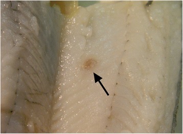
The European perch, Perca fluviatilis L. is a common paratenic host of dioctophymatid nematodes belonging to the genus Eustrongylides. In this host, once infected oligochaetes, which serve as the first intermediate host, are ingested, Eustrongylides migrates through the intestine and is frequently encountered within the musculature, free within the body cavity, or encapsulated on the viscera. The current study details the first Italian record of Eustrongylides sp. with larvae reported in the muscle of P. fluviatilis.
Uninfected and nematode-infected muscle tissues of perch were fixed and prepared for histological evaluation and electron microscopy. Some sections were subjected to an indirect immunohistochemical method using anti-PCNA, anti-piscidin 3 and anti-piscidin 4 antibodies.
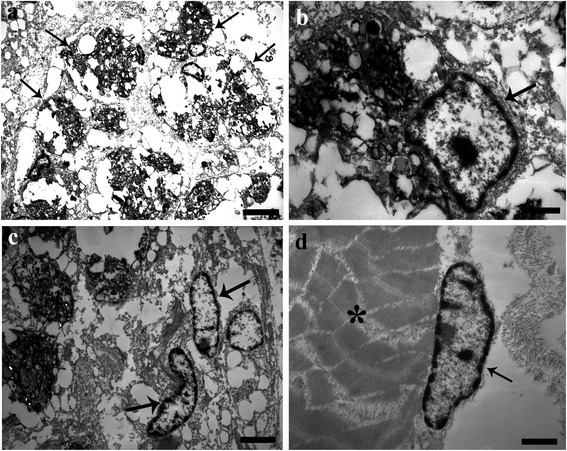
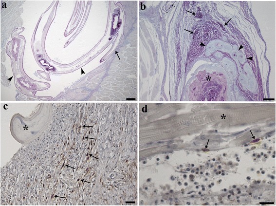
A total of 510 P. fluviatilis (TL range 15-25 cm) from Lake Trasimeno, Perugia were post-mortemed; 31 individuals had encysted nematode larvae within their musculature (1-2 worms fish(-1)). Histologically, larvae were surrounded by a capsule with an evident acute inflammatory reaction. Muscle degeneration and necrosis extending throughout the sarcoplasm, sarcolemmal basal lamina, endomysial connective tissue cells and capillaries was frequently observed. Within the encapsulating reaction, macrophage aggregates (MAs) were seen. Immunohistochemical staining with the proliferating cell nuclear antigen (PCNA) revealed numerous PCNA-positive cells within the thickness of the capsule and in the immediate vicinity surrounding Eustrongylides sp. larvae (i.e. fibroblasts and satellite cells), suggesting a host response had been initiated to repair the nematode-damaged muscle. Mast cells (MCs) staining positively for piscidin 3, were demonstrated for the first time in response to a muscle-infecting nematode. The piscidin 3 positive MC's were seen principally in the periphery of the capsule surrounding the Eustrongylides sp. larva.
A host tissue response to Eustrongylides sp. larvae infecting the musculature of P. fluviatilis was observed. Numerous fibroblasts, MAs and MCs were seen throughout the thick fibroconnectival layer of the capsule enclosing larvae. PCNA positive cells within the capsule suggest that host repair of nematode damaged muscle does occur, while the presence of the antimicrobial peptide piscidin 3 is shown for the first time. This is first report of Eustrongylides sp. in an Italian population of P. fluviatilis.
欧洲鲈(Perca fluviatilis L.)是属于真圆线虫属的膨结线虫的常见转续宿主。在这种宿主体内,一旦摄入作为第一中间宿主的感染性寡毛类动物,真圆线虫就会穿过肠道,经常在肌肉组织中、体腔内自由存在或包裹在内脏上被发现。本研究详细记录了意大利首次在欧洲鲈肌肉中发现带有幼虫的真圆线虫属的情况。
将未感染和感染线虫的鲈鱼肌肉组织固定并制备用于组织学评估和电子显微镜检查。一些切片采用抗增殖细胞核抗原(PCNA)、抗鱼杀菌肽3和抗鱼杀菌肽4抗体进行间接免疫组织化学方法检测。
对来自佩鲁贾特拉西梅诺湖的510条欧洲鲈(全长范围为15 - 25厘米)进行了死后检查;31条个体的肌肉组织中有包囊化的线虫幼虫(每条鱼1 - 2条虫)。组织学上,幼虫被一个有明显急性炎症反应的包囊包围。经常观察到肌肉变性和坏死延伸至整个肌浆、肌膜基底层、肌内膜结缔组织细胞和毛细血管。在包囊反应中可见巨噬细胞聚集物(MAs)。用增殖细胞核抗原(PCNA)进行免疫组织化学染色显示,在包囊厚度内以及真圆线虫属幼虫紧邻区域(即成纤维细胞和卫星细胞)有大量PCNA阳性细胞,表明宿主已启动修复线虫损伤肌肉的反应。首次证明了对感染肌肉的线虫有鱼杀菌肽3阳性的肥大细胞(MCs)。鱼杀菌肽3阳性的肥大细胞主要见于包围真圆线虫属幼虫的包囊周边。
观察到宿主组织对感染欧洲鲈肌肉组织的真圆线虫属幼虫的反应。在包围幼虫的包囊厚纤维结缔组织层中可见大量成纤维细胞、巨噬细胞聚集物和肥大细胞。包囊内PCNA阳性细胞表明宿主确实对线虫损伤的肌肉进行了修复,同时首次显示了抗菌肽鱼杀菌肽3的存在。这是意大利欧洲鲈种群中首次报道真圆线虫属。