• 文献检索
  • 文档翻译
  • 深度研究
  • 学术资讯
  • Suppr Zotero 插件Zotero 插件
  • 邀请有礼
  • 套餐&价格
  • 历史记录
应用&插件
Suppr Zotero 插件Zotero 插件浏览器插件Mac 客户端Windows 客户端微信小程序
定价
高级版会员购买积分包购买API积分包
服务
文献检索文档翻译深度研究API 文档MCP 服务
关于我们
关于 Suppr公司介绍联系我们用户协议隐私条款
关注我们

Suppr 超能文献

核心技术专利:CN118964589B侵权必究
粤ICP备2023148730 号-1Suppr @ 2026

文献检索

告别复杂PubMed语法,用中文像聊天一样搜索,搜遍4000万医学文献。AI智能推荐,让科研检索更轻松。

立即免费搜索

文件翻译

保留排版,准确专业,支持PDF/Word/PPT等文件格式,支持 12+语言互译。

免费翻译文档

深度研究

AI帮你快速写综述,25分钟生成高质量综述,智能提取关键信息,辅助科研写作。

立即免费体验

相似文献

1
Reducing Neointima Formation in a Swine Model with IVUS and Sirolimus Microbubbles.使用血管内超声和西罗莫司微泡减少猪模型中的新生内膜形成。
Ann Biomed Eng. 2015 Nov;43(11):2642-51. doi: 10.1007/s10439-015-1315-6. Epub 2015 Apr 17.
2
Paclitaxcel-coated balloon plus bare metal stent vs. sirolimus-eluting stent in de novo lesions: an IVUS study.紫杉醇涂层球囊联合裸金属支架与雷帕霉素洗脱支架治疗初发病变:一项血管内超声研究。
EuroIntervention. 2012 Aug;8(4):450-5. doi: 10.4244/EIJV8I4A71.
3
Local delivery of sirolimus nanoparticles for the treatment of in-stent restenosis.局部递送西罗莫司纳米粒治疗支架内再狭窄。
Catheter Cardiovasc Interv. 2013 Feb;81(2):E124-9. doi: 10.1002/ccd.24331. Epub 2012 May 4.
4
Intravascular ultrasound results from the NEVO ResElution-I trial: a randomized, blinded comparison of sirolimus-eluting NEVO stents with paclitaxel-eluting TAXUS Liberté stents in de novo native coronary artery lesions.NEVO ResElution-I 试验的血管内超声结果:在新出现的原生冠状动脉病变中,雷帕霉素洗脱 NEVO 支架与紫杉醇洗脱 TAXUS Liberté 支架的随机、盲法比较。
Circ Cardiovasc Interv. 2011 Apr 1;4(2):146-54. doi: 10.1161/CIRCINTERVENTIONS.110.957175. Epub 2011 Mar 8.
5
Optimal stent-sizing with intravascular ultrasound contributes to complete neointimal coverage after sirolimus-eluting stent implantation assessed by angioscopy.血管内超声指导的最佳支架尺寸选择有助于经血管内超声评估的西罗莫司洗脱支架置入后完全的新生内膜覆盖。
JACC Cardiovasc Interv. 2009 Oct;2(10):989-94. doi: 10.1016/j.jcin.2009.07.006.
6
Novel site-specific systemic delivery of Rapamycin with perfluorobutane gas microbubble carrier reduced neointimal formation in a porcine coronary restenosis model.使用全氟丁烷气体微泡载体进行雷帕霉素的新型位点特异性全身递送可减少猪冠状动脉再狭窄模型中的内膜增生。
Catheter Cardiovasc Interv. 2005 Mar;64(3):389-94. doi: 10.1002/ccd.20285.
7
Segmental vessel wall shear stress and neointimal formation after sirolimus-eluting stent implantation: physiological insights in a porcine coronary model.西罗莫司洗脱支架植入术后节段性血管壁剪切应力与新生内膜形成:猪冠状动脉模型的生理学见解
Cardiovasc Revasc Med. 2005 Apr-Jun;6(2):58-64. doi: 10.1016/j.carrev.2005.05.004.
8
Sirolimus-eluting stent implantation in small coronary arteries: a three dimensional intravascular ultrasound study from the SIRIUS trial.西罗莫司洗脱支架置入小冠状动脉:来自 SIRIUS 试验的三维血管内超声研究。
Int J Cardiol. 2010 Jan 21;138(2):126-30. doi: 10.1016/j.ijcard.2008.08.006. Epub 2008 Sep 19.
9
Negative vascular remodelling after implantation of bioabsorbable magnesium alloy stents in porcine coronary arteries: a randomised comparison with bare-metal and sirolimus-eluting stents.生物可吸收镁合金支架植入猪冠状动脉后的负性血管重塑:与裸金属支架和西罗莫司洗脱支架的随机对照比较
Heart. 2009 Mar;95(3):241-6. doi: 10.1136/hrt.2007.139261. Epub 2008 Aug 22.
10
Sirolimus-loaded stealth colloidal systems attenuate neointimal hyperplasia after balloon injury: a comparison of phospholipid micelles and liposomes.载西罗莫司隐形胶态系统减轻球囊损伤后内膜增生:磷脂胶束与脂质体的比较。
Int J Pharm. 2013 Oct 15;455(1-2):320-30. doi: 10.1016/j.ijpharm.2013.07.003. Epub 2013 Jul 16.

引用本文的文献

1
Focused Ultrasound: Noninvasive Image-Guided Therapy.聚焦超声:非侵入性图像引导治疗。
Invest Radiol. 2025 Mar 1;60(3):205-219. doi: 10.1097/RLI.0000000000001116. Epub 2024 Aug 21.
2
Ultrasonic technologies in imaging and drug delivery.超声技术在成像和药物传递中的应用。
Cell Mol Life Sci. 2021 Sep;78(17-18):6119-6141. doi: 10.1007/s00018-021-03904-9. Epub 2021 Jul 23.
3
Ultrasound-Responsive Cavitation Nuclei for Therapy and Drug Delivery.用于治疗和药物递送的超声响应性空化核
Ultrasound Med Biol. 2020 Jun;46(6):1296-1325. doi: 10.1016/j.ultrasmedbio.2020.01.002. Epub 2020 Mar 10.
4
Endurance exercise training does not limit coronary atherosclerosis in familial hypercholesterolemic swine.耐力运动训练并不能限制家族性高胆固醇血症猪的冠状动脉粥样硬化。
Physiol Rep. 2019 Feb;7(4):e14008. doi: 10.14814/phy2.14008.
5
Efficacy of Sonothrombolysis Using Microbubbles Produced by a Catheter-Based Microfluidic Device in a Rat Model of Ischemic Stroke.基于导管微流控装置产生的微泡的声溶栓在大鼠缺血性脑卒中模型中的疗效。
Ann Biomed Eng. 2019 Apr;47(4):1012-1022. doi: 10.1007/s10439-019-02209-0. Epub 2019 Jan 28.
6
Rapamycin: A Bacteria-Derived Immunosuppressant That Has Anti-atherosclerotic Effects and Its Clinical Application.雷帕霉素:一种具有抗动脉粥样硬化作用的细菌衍生免疫抑制剂及其临床应用。
Front Pharmacol. 2019 Jan 7;9:1520. doi: 10.3389/fphar.2018.01520. eCollection 2018.

本文引用的文献

1
An IVUS transducer for microbubble therapies.用于微泡治疗的血管内超声换能器。
IEEE Trans Ultrason Ferroelectr Freq Control. 2014 Mar;61(3):441-9. doi: 10.1109/TUFFC.2014.2929.
2
Heart disease and stroke statistics--2014 update: a report from the American Heart Association.《2014年心脏病和中风统计数据更新:美国心脏协会报告》
Circulation. 2014 Jan 21;129(3):e28-e292. doi: 10.1161/01.cir.0000441139.02102.80. Epub 2013 Dec 18.
3
Relationship between intravascular ultrasound guidance and clinical outcomes after drug-eluting stents: the assessment of dual antiplatelet therapy with drug-eluting stents (ADAPT-DES) study.血管内超声指导与药物洗脱支架治疗后临床结局的关系:药物洗脱支架双重抗血小板治疗评估(ADAPT-DES)研究。
Circulation. 2014 Jan 28;129(4):463-70. doi: 10.1161/CIRCULATIONAHA.113.003942. Epub 2013 Nov 26.
4
Are the culprit lesions severely stenotic?罪魁祸首病变是否严重狭窄?
JACC Cardiovasc Imaging. 2013 Oct;6(10):1108-1114. doi: 10.1016/j.jcmg.2013.05.004.
5
Enhanced intracellular delivery of a model drug using microbubbles produced by a microfluidic device.利用微流控装置产生的微泡增强模型药物的细胞内递送。
Ultrasound Med Biol. 2013 Jul;39(7):1267-76. doi: 10.1016/j.ultrasmedbio.2013.01.023. Epub 2013 Apr 30.
6
Contemporary patterns of fractional flow reserve and intravascular ultrasound use among patients undergoing percutaneous coronary intervention in the United States: insights from the National Cardiovascular Data Registry.美国接受经皮冠状动脉介入治疗患者中血流储备分数和血管内超声的当代使用模式:来自国家心血管数据注册库的见解
J Am Coll Cardiol. 2012 Dec 4;60(22):2337-9. doi: 10.1016/j.jacc.2012.08.990.
7
Intravascular ultrasound catheter to enhance microbubble-based drug delivery via acoustic radiation force.血管内超声导管增强基于微泡的药物输送的声辐射力。
IEEE Trans Ultrason Ferroelectr Freq Control. 2012 Oct;59(10):2156-66. doi: 10.1109/TUFFC.2012.2442.
8
Spatiotemporally controlled single cell sonoporation.时空控制的单细胞声孔法。
Proc Natl Acad Sci U S A. 2012 Oct 9;109(41):16486-91. doi: 10.1073/pnas.1208198109. Epub 2012 Sep 24.
9
Focused ultrasound-mediated drug delivery from microbubbles reduces drug dose necessary for therapeutic effect on neointima formation--brief report.聚焦超声介导微泡载药减少了治疗新生内膜形成所需的药物剂量——简短报告。
Arterioscler Thromb Vasc Biol. 2011 Dec;31(12):2853-5. doi: 10.1161/ATVBAHA.111.238170. Epub 2011 Sep 29.
10
The impact of patient and lesion complexity on clinical and angiographic outcomes after revascularization with zotarolimus- and everolimus-eluting stents: a substudy of the RESOLUTE All Comers Trial (a randomized comparison of a zotarolimus-eluting stent with an everolimus-eluting stent for percutaneous coronary intervention).在接受依维莫司洗脱支架和佐他莫司洗脱支架血运重建后的患者和病变复杂性对临床和血管造影结果的影响:RESOLUTE 所有患者试验的亚研究(依维莫司洗脱支架与佐他莫司洗脱支架经皮冠状动脉介入治疗的随机比较)。
J Am Coll Cardiol. 2011 May 31;57(22):2221-32. doi: 10.1016/j.jacc.2011.01.036.

使用血管内超声和西罗莫司微泡减少猪模型中的新生内膜形成。

Reducing Neointima Formation in a Swine Model with IVUS and Sirolimus Microbubbles.

作者信息

Kilroy Joseph P, Dhanaliwala Ali H, Klibanov Alexander L, Bowles Douglas K, Wamhoff Brian R, Hossack John A

机构信息

Department of Biomedical Engineering, University of Virginia, Charlottesville, VA, 22908, USA.

School of Medicine, University of Virginia, Charlottesville, VA, 22908, USA.

出版信息

Ann Biomed Eng. 2015 Nov;43(11):2642-51. doi: 10.1007/s10439-015-1315-6. Epub 2015 Apr 17.

DOI:10.1007/s10439-015-1315-6
PMID:25893508
原文链接:https://pmc.ncbi.nlm.nih.gov/articles/PMC4609231/
Abstract

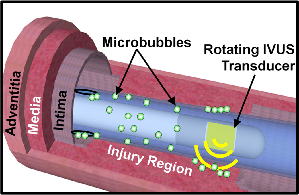
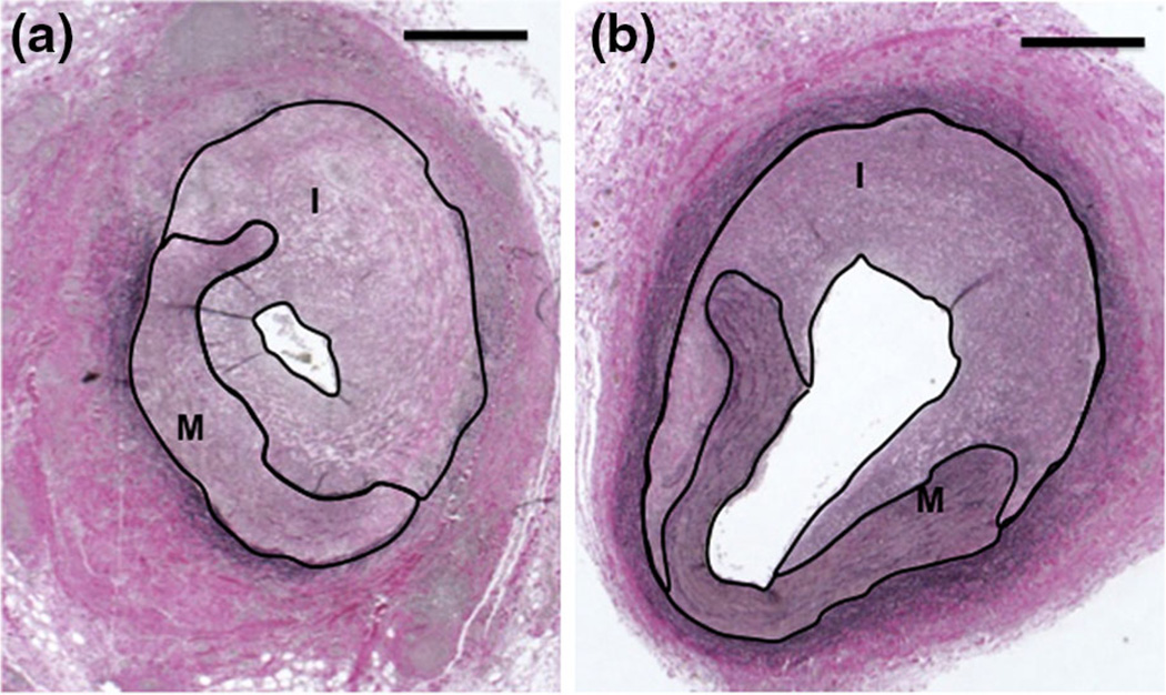
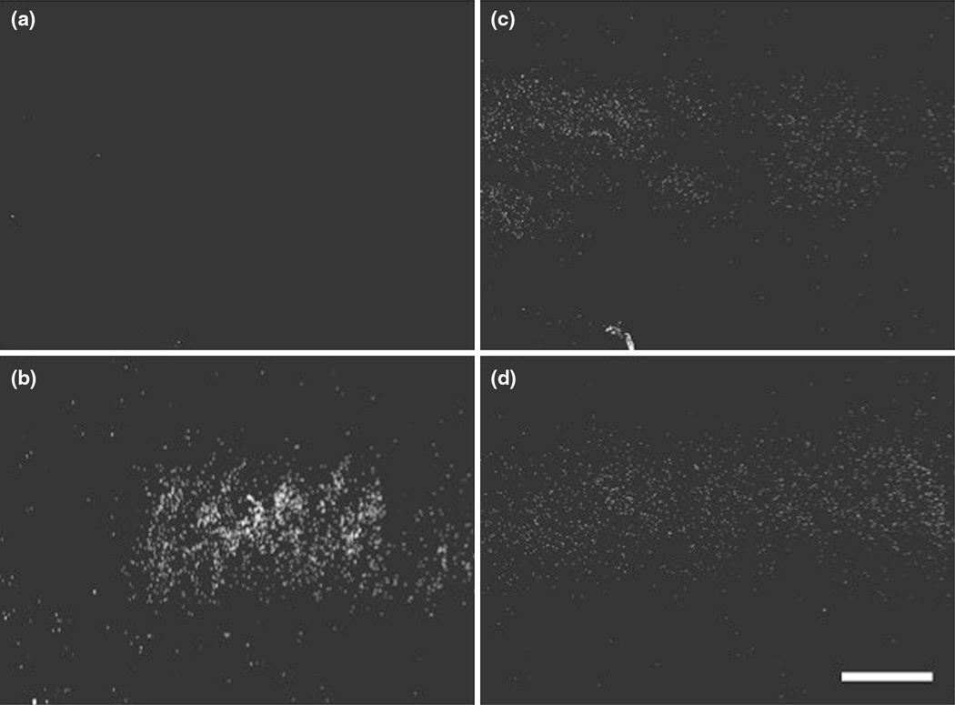
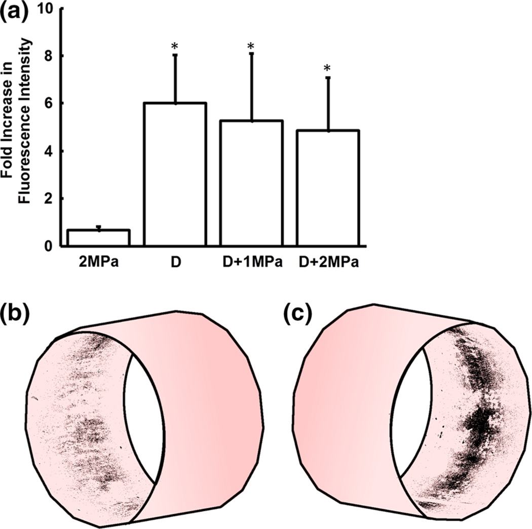
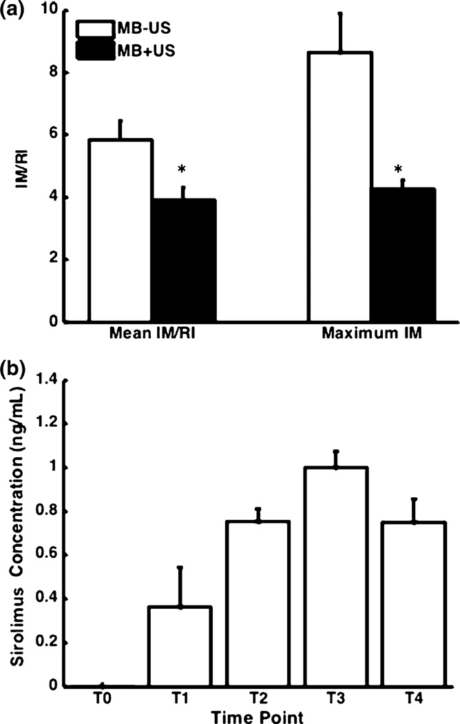
Potent therapeutic compounds with dose dependent side effects require more efficient and selective drug delivery to reduce systemic drug doses. Here, we demonstrate a new platform that combines intravascular ultrasound (IVUS) and drug-loaded microbubbles to enhance and localize drug delivery, while enabling versatility of drug type and dosing. Localization and degree of delivery with IVUS and microbubbles was assessed using fluorophore-loaded microbubbles and different IVUS parameters in ex vivo swine arteries. Using a swine model of neointimal hyperplasia, reduction of neointima formation following balloon injury was evaluated when using the combination of IVUS and sirolimus-loaded microbubbles. IVUS and microbubble enhanced fluorophore delivery was greatest when applying low amplitude pulses in the ex vivo model. In the in vivo model, neointima formation was reduced by 50% after treatment with IVUS and the sirolimus-loaded microbubbles. This reduction was achieved with a sirolimus whole blood concentration comparable to a commercial drug-eluting stent (0.999 ng/mL). We anticipate this therapy will find clinical use localizing drug delivery for numerous other diseases in addition to serving as an adjunct to stents in treating atherosclerosis.

摘要

具有剂量依赖性副作用的强效治疗化合物需要更高效、更具选择性的药物递送方式,以降低全身药物剂量。在此,我们展示了一个新平台,该平台将血管内超声(IVUS)与载药微泡相结合,既能增强药物递送并使其定位,又能实现药物类型和给药方式的多样化。在离体猪动脉中,使用载有荧光团的微泡和不同的IVUS参数评估了IVUS和微泡的定位及递送程度。利用猪内膜增生模型,评估了在使用IVUS与载西罗莫司微泡联合治疗时,球囊损伤后内膜增生的减少情况。在离体模型中,施加低振幅脉冲时,IVUS和微泡增强的荧光团递送效果最佳。在体内模型中,用IVUS和载西罗莫司微泡治疗后,内膜增生减少了50%。实现这种减少所需要的西罗莫司全血浓度与商用药物洗脱支架相当(0.999纳克/毫升)。我们预计,这种疗法除了可作为治疗动脉粥样硬化时支架的辅助手段外,还将在定位药物递送治疗众多其他疾病方面得到临床应用。

https://cdn.ncbi.nlm.nih.gov/pmc/blobs/8f98/4609231/0a689beaf104/nihms682101f5.jpg
https://cdn.ncbi.nlm.nih.gov/pmc/blobs/8f98/4609231/96ba971539a5/nihms682101f1.jpg
https://cdn.ncbi.nlm.nih.gov/pmc/blobs/8f98/4609231/1cf2e9729a72/nihms682101f2.jpg
https://cdn.ncbi.nlm.nih.gov/pmc/blobs/8f98/4609231/4f02faf0aa56/nihms682101f3.jpg
https://cdn.ncbi.nlm.nih.gov/pmc/blobs/8f98/4609231/ad4430bdab18/nihms682101f4.jpg
https://cdn.ncbi.nlm.nih.gov/pmc/blobs/8f98/4609231/0a689beaf104/nihms682101f5.jpg