Khairoun Meriem, van den Heuvel Mieke, van den Berg Bernard M, Sorop Oana, de Boer Rients, van Ditzhuijzen Nienke S, Bajema Ingeborg M, Baelde Hans J, Zandbergen Malu, Duncker Dirk J, Rabelink Ton J, Reinders Marlies E J, van der Giessen Wim J, Rotmans Joris I
Department of Nephrology, Leiden University Medical Center, Leiden, The Netherlands.
Division of Experimental Cardiology, Department of Cardiology, Erasmus Medical Center Rotterdam, Rotterdam, The Netherlands.
PLoS One. 2015 Apr 24;10(4):e0121555. doi: 10.1371/journal.pone.0121555. eCollection 2015.
Diabetes mellitus (DM) is associated with a range of microvascular complications including diabetic nephropathy (DN). Microvascular abnormalities in the kidneys are common histopathologic findings in DN, which represent one manifestation of ongoing systemic microvascular damage. Recently, sidestream dark-field (SDF) imaging has emerged as a noninvasive tool that enables one to visualize the microcirculation. In this study, we investigated whether changes in the systemic microvasculature induced by DM and an atherogenic diet correlated spatiotemporally with renal damage.
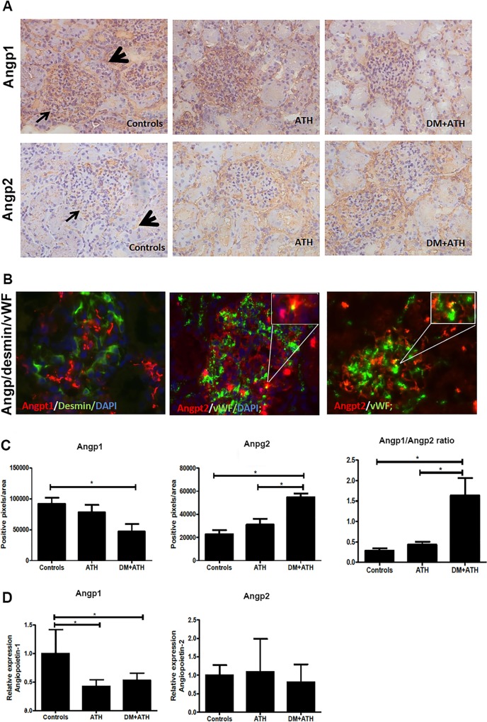
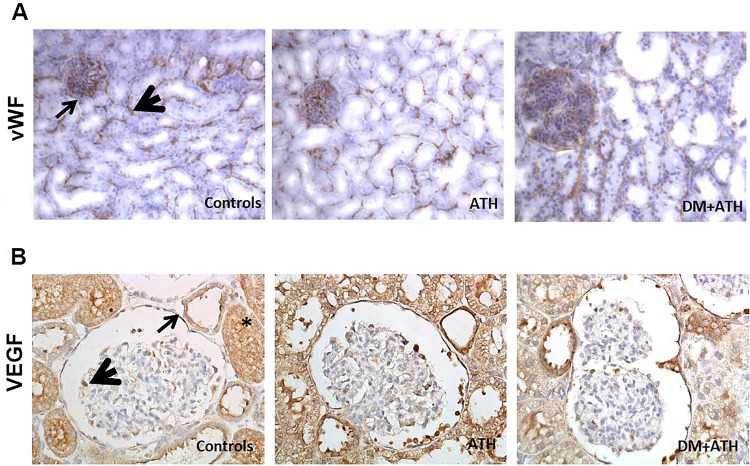
Atherosclerotic lesion development was triggered in streptozotocin-induced DM pigs (140 mg/kg body weight) by administering an atherogenic diet for approximately 11 months. Fifteen months following induction of DM, microvascular morphology was visualized in control pigs (n = 7), non-diabetic pigs fed an atherogenic diet (ATH, n = 5), and DM pigs fed an atherogenic diet (DM+ATH, n = 5) using SDF imaging of oral mucosal tissue. Subsequently, kidneys were harvested from anethesized pigs and the expression levels of well-established markers for microvascular integrity, such as Angiopoietin-1 (Angpt1) and Angiopoietin-2 (Angpt2) were determined immunohistochemically, while endothelial cell (EC) abundance was determined by immunostaining for von Willebrand factor (vWF).
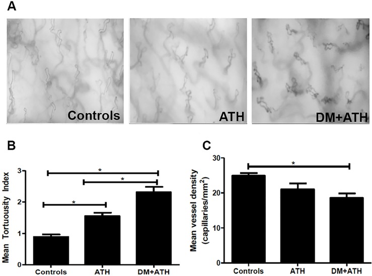
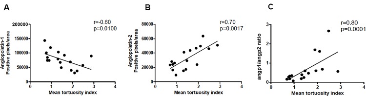
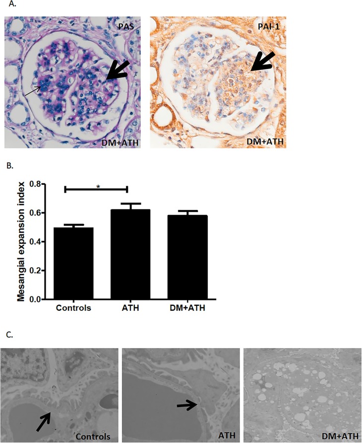
Our study revealed an increase in the capillary tortuosity index in DM+ATH pigs (2.31±0.17) as compared to the control groups (Controls 0.89±0.08 and ATH 1.55±0.11; p<0.05). Kidney biopsies showed marked glomerular lesions consisting of mesangial expansion and podocyte lesions. Furthermore, we observed a disturbed Angpt2/Angpt1 balance in the cortex of the kidney, as evidenced by increased expression of Angpt2 in DM+ATH pigs as compared to Control pigs (p<0.05).
In the setting of DM, atherogenesis leads to the augmentation of mucosal capillary tortuosity, indicative of systemic microvascular damage. Concomitantly, a dysbalance in renal angiopoietins was correlated with the development of diabetic nephropathy. As such, our studies strongly suggest that defects in the systemic microvasculature mirror the accumulation of microvascular damage in the kidney.
糖尿病(DM)与一系列微血管并发症相关,包括糖尿病肾病(DN)。肾脏微血管异常是DN常见的组织病理学表现,代表了全身性微血管持续损伤的一种表现形式。最近,侧流暗视野(SDF)成像已成为一种非侵入性工具,能够使人们可视化微循环。在本研究中,我们调查了DM和致动脉粥样硬化饮食诱导的全身微血管变化是否与肾损伤在时空上相关。
通过给予致动脉粥样硬化饮食约11个月,在链脲佐菌素诱导的DM猪(140mg/kg体重)中引发动脉粥样硬化病变发展。DM诱导15个月后,使用口腔黏膜组织的SDF成像观察对照猪(n = 7)、喂食致动脉粥样硬化饮食的非糖尿病猪(ATH,n = 5)和喂食致动脉粥样硬化饮食的DM猪(DM+ATH,n = 5)的微血管形态。随后,从麻醉的猪身上采集肾脏,免疫组织化学测定微血管完整性的成熟标志物如血管生成素-1(Angpt1)和血管生成素-2(Angpt2)的表达水平,而通过对血管性血友病因子(vWF)进行免疫染色来确定内皮细胞(EC)丰度。
我们的研究显示,与对照组相比,DM+ATH猪的毛细血管迂曲指数增加(2.31±0.17)(对照组为0.89±0.08,ATH组为1.55±0.11;p<0.05)。肾脏活检显示明显的肾小球病变,包括系膜扩张和足细胞病变。此外,我们观察到肾脏皮质中Angpt2/Angpt1平衡紊乱,与对照猪相比,DM+ATH猪中Angpt2表达增加证明了这一点(p<0.05)。
在DM情况下,动脉粥样硬化导致黏膜毛细血管迂曲增加,表明全身性微血管损伤。同时,肾脏血管生成素失衡与糖尿病肾病的发展相关。因此,我们的研究强烈表明全身微血管缺陷反映了肾脏微血管损伤的积累。