Hänggi Jürgen, Langer Nicolas, Lutz Kai, Birrer Karin, Mérillat Susan, Jäncke Lutz
Division Neuropsychology, Department of Psychology, University of Zurich, Zurich, Switzerland.
Division Neuropsychology, Department of Psychology, University of Zurich, Zurich, Switzerland; Neural Systems Lab, The City College of New York, New York, NY, United States of America; Child Mind Institute, New York, NY, United States of America.
PLoS One. 2015 Apr 27;10(4):e0124222. doi: 10.1371/journal.pone.0124222. eCollection 2015.
There is no doubt that good bimanual performance is very important for skilled handball playing. The control of the non-dominant hand is especially demanding since efficient catching and throwing needs both hands.
METHODOLOGY/HYPOTHESES: We investigated training-induced structural neuroplasticity in professional handball players using several structural neuroimaging techniques and analytic approaches and also provide a review of the literature about sport-induced structural neuroplastic alterations. Structural brain adaptations were expected in regions relevant for motor and somatosensory processing such as the grey matter (GM) of the primary/secondary motor (MI/supplementary motor area, SMA) and somatosensory cortex (SI/SII), basal ganglia, thalamus, and cerebellum and in the white matter (WM) of the corticospinal tract (CST) and corpus callosum, stronger in brain regions controlling the non-dominant left hand.
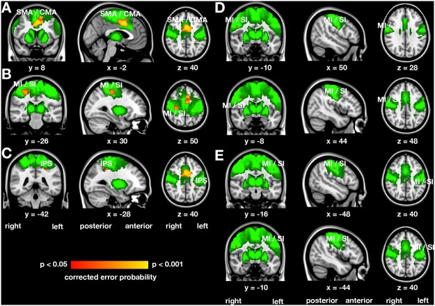
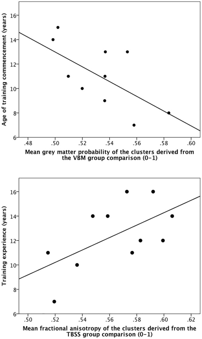
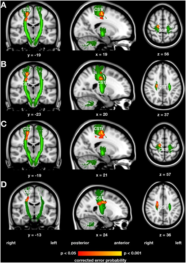
Increased GM volume in handball players compared with control subjects were found in the right MI/SI, bilateral SMA/cingulate motor area, and left intraparietal sulcus. Fractional anisotropy (FA) and axial diffusivity were increased within the right CST in handball players compared with control women. Age of handball training commencement correlated inversely with GM volume in the right and left MI/SI and years of handball training experience correlated inversely with radial diffusivity in the right CST. Subcortical structures tended to be larger in handball players. The anatomical measures of the brain regions associated with handball playing were positively correlated in handball players, but not interrelated in control women.
DISCUSSION/CONCLUSION: Training-induced structural alterations were found in the somatosensory-motor network of handball players, more pronounced in the right hemisphere controlling the non-dominant left hand. Correlations between handball training-related measures and anatomical differences suggest neuroplastic adaptations rather than a genetic predisposition for a ball playing affinity. Investigations of neuroplasticity specifically in sportsmen might help to understand the neural mechanisms of expertise in general.
毫无疑问,良好的双手协调性对于熟练地进行手球运动非常重要。非优势手的控制尤其具有挑战性,因为高效的接球和投球需要双手配合。
方法/假设:我们使用多种结构神经成像技术和分析方法,研究了职业手球运动员训练诱导的结构神经可塑性,并对运动诱导的结构神经可塑性改变的文献进行了综述。预计在与运动和体感处理相关的区域会出现大脑结构适应性变化,如初级/次级运动区(MI/辅助运动区,SMA)和体感皮层(SI/SII)、基底神经节、丘脑和小脑的灰质(GM),以及皮质脊髓束(CST)和胼胝体的白质(WM),在控制非优势左手的脑区变化更明显。
与对照组相比,手球运动员右侧MI/SI、双侧SMA/扣带运动区和左侧顶内沟的GM体积增加。与对照女性相比,手球运动员右侧CST内的分数各向异性(FA)和轴向扩散率增加。手球训练开始的年龄与左右MI/SI的GM体积呈负相关,手球训练经验的年限与右侧CST的径向扩散率呈负相关。手球运动员的皮层下结构往往更大。与手球运动相关的脑区解剖学测量值在运动员中呈正相关,但在对照女性中无相关性。
讨论/结论:在手球运动员的体感运动网络中发现了训练诱导的结构改变,在控制非优势左手的右半球更为明显。手球训练相关指标与解剖学差异之间的相关性表明是神经可塑性适应,而非对球类运动的遗传易感性。专门针对运动员的神经可塑性研究可能有助于一般地理解专业技能的神经机制。