Lam Marnix G E H, Banerjee Arjun, Goris Michael L, Iagaru Andrei H, Mittra Erik S, Louie John D, Sze Daniel Y
Division of Interventional Radiology, Stanford University, Stanford, CA, USA,
Eur J Nucl Med Mol Imaging. 2015 Jul;42(8):1192-201. doi: 10.1007/s00259-015-3048-z. Epub 2015 Apr 28.
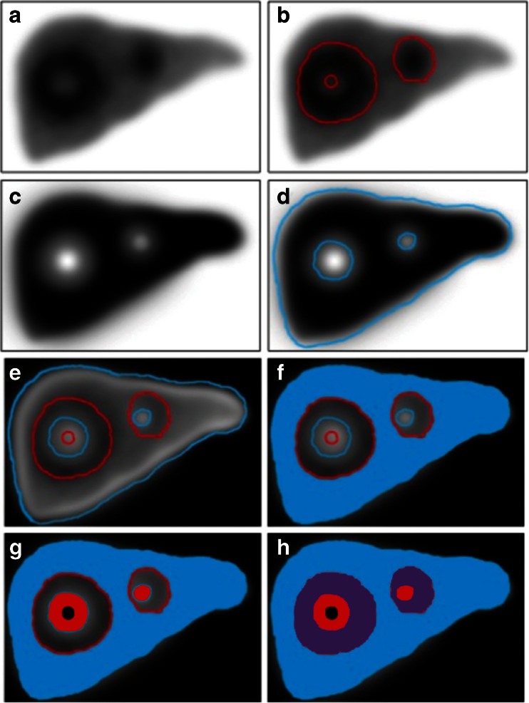
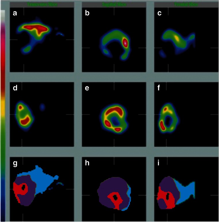
Fusion dual-tracer SPECT imaging enables physiological rather than morphological voxel-based partitioning and dosimetry for (90)Y hepatic radioembolization (RE). We evaluated its prognostic value in a large heterogeneous cohort of patients with extensive hepatic malignancy.
A total of 122 patients with primary or secondary liver malignancy (18 different cell types) underwent SPECT imaging after intraarterial injection of (99m)Tc macroaggregated albumin (TcMAA) as a simulation of subsequent (90)Y microsphere distribution, followed by administration of an excess of intravenous (99m)Tc-labelled sulphur colloid (TcSC) as a biomarker for functional liver, and a second SPECT scan. TcMAA distribution was used to estimate (90)Y radiation absorbed dose in tumour (D T) and in functional liver. Laboratory and clinical follow-up were recorded for 12 weeks after RE, and radiographic responses according to (m)RECIST were evaluated at 3 and 6 months. Dose-response relationships were determined for efficacy and toxicity.
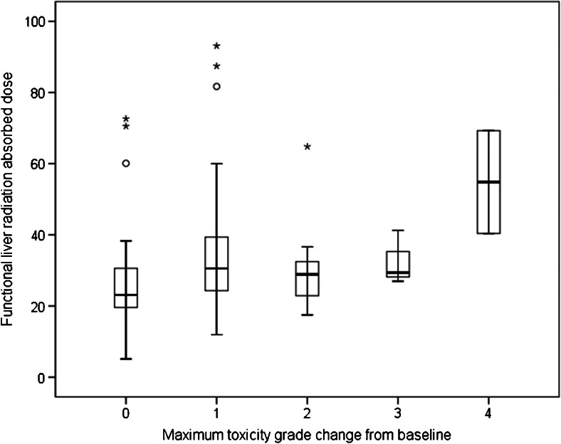
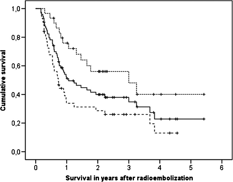
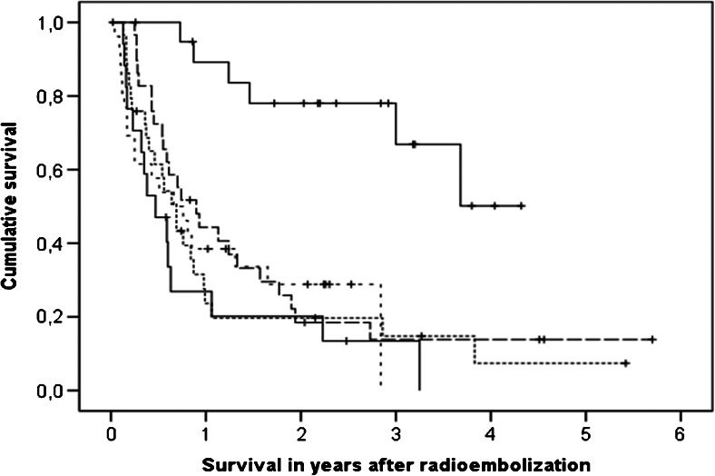
Patients were treated with a median of 1.73 GBq activity of resin microspheres (98 patients) or glass microspheres (24 patients), in a whole-liver approach (97 patients) or a lobar approach (25 patients). The objective response rate was 41% at 3 months and 48% at 6 months. Response was correlated with D T (P < 0.01). Median overall survival was 10.1 months (95% confidence interval 7.4 - 12.8 months). Responders lived for 36.0 months compared to 8.7 months for nonresponders (P < 0.01). Stratified for tumour cell type, D T was independently associated with survival (P < 0.01). Absorbed dose in functional liver was correlated with toxicity grade change (P < 0.05) and RE-induced liver disease (P < 0.05).
Fusion dual-tracer SPECT imaging offers a physiology-based functional imaging tool to predict efficacy and toxicity of RE. This technique can be refined to define dosing thresholds for specific tumour types and treatments, but appears generally predictive even in a heterogeneous cohort.
融合双示踪剂单光子发射计算机断层扫描(SPECT)成像可为钇-90(90Y)肝动脉栓塞术(RE)提供基于生理学而非形态学的体素分割和剂量测定。我们在一大组异质性广泛肝恶性肿瘤患者中评估了其预后价值。
总共122例原发性或继发性肝恶性肿瘤患者(18种不同细胞类型)在动脉内注射锝-99m(99mTc)标记的大颗粒白蛋白(TcMAA)以模拟随后的90Y微球分布后接受SPECT成像,随后给予过量静脉注射的99mTc标记的硫胶体(TcSC)作为功能性肝脏的生物标志物,并进行第二次SPECT扫描。TcMAA分布用于估计肿瘤(DT)和功能性肝脏中的90Y辐射吸收剂量。RE后12周记录实验室和临床随访情况,并在3个月和6个月时根据改良实体瘤疗效评价标准(mRECIST)评估影像学反应。确定疗效和毒性的剂量反应关系。
患者接受了中位活度为1.73GBq的树脂微球(98例)或玻璃微球(24例)治疗,采用全肝治疗方法(97例)或叶治疗方法(25例)。3个月时客观缓解率为41%,6个月时为48%。缓解与DT相关(P<0.01)。中位总生存期为10.1个月(95%置信区间7.4 - 12.8个月)。缓解者生存36.0个月,未缓解者生存8.7个月(P<0.01)。按肿瘤细胞类型分层,DT与生存独立相关(P<0.01)。功能性肝脏中的吸收剂量与毒性分级变化(P<0.05)和RE诱导的肝病(P<0.05)相关。
融合双示踪剂SPECT成像提供了一种基于生理学的功能成像工具,可预测RE的疗效和毒性。该技术可进一步完善以确定特定肿瘤类型和治疗的给药阈值,但即使在异质性队列中似乎也具有普遍预测性。