Bansal Aditya, Pandey Mukesh K, Demirhan Yunus E, Nesbitt Jonathan J, Crespo-Diaz Ruben J, Terzic Andre, Behfar Atta, DeGrado Timothy R
Department of Radiology, Mayo Clinic, Rochester, 55905 MN USA.
Division of Cardiovascular Diseases, Mayo Clinic, Rochester, 55905 MN USA.
EJNMMI Res. 2015 Mar 28;5:19. doi: 10.1186/s13550-015-0098-y. eCollection 2015.
With the recent growth of interest in cell-based therapies and radiolabeled cell products, there is a need to develop more robust cell labeling and imaging methods for in vivo tracking of living cells. This study describes evaluation of a novel cell labeling approach with the positron emission tomography (PET) isotope (89)Zr (T 1/2 = 78.4 h). (89)Zr may allow PET imaging measurements for several weeks and take advantage of the high sensitivity of PET imaging.
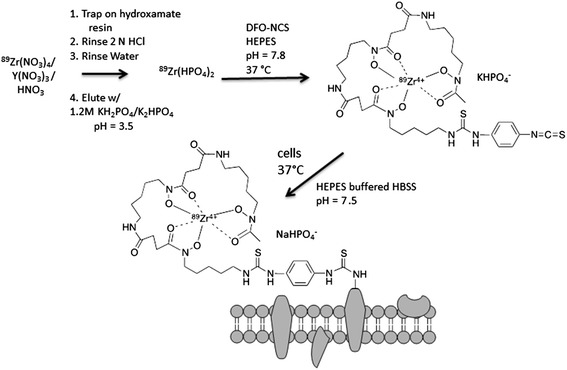
A novel cell labeling agent, (89)Zr-desferrioxamine-NCS ((89)Zr-DBN), was synthesized. Mouse-derived melanoma cells (mMCs), dendritic cells (mDCs), and human mesenchymal stem cells (hMSCs) were covalently labeled with (89)Zr-DBN via the reaction between the NCS group on (89)Zr-DBN and primary amine groups present on cell surface membrane protein. The stability of the label on the cell was tested by cell efflux studies for 7 days. The effect of labeling on cellular viability was tested by proliferation, trypan blue, and cytotoxicity/apoptosis assays. The stability of label was also studied in in vivo mouse models by serial PET scans and ex vivo biodistribution following intravenous and intramyocardial injection of (89)Zr-labeled hMSCs. For comparison, imaging experiments were performed after intravenous injections of (89)Zr hydrogen phosphate ((89)Zr(HPO4)2).
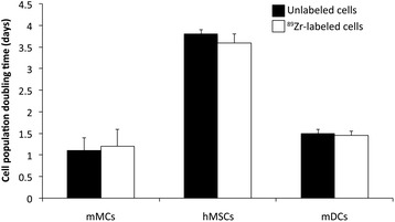
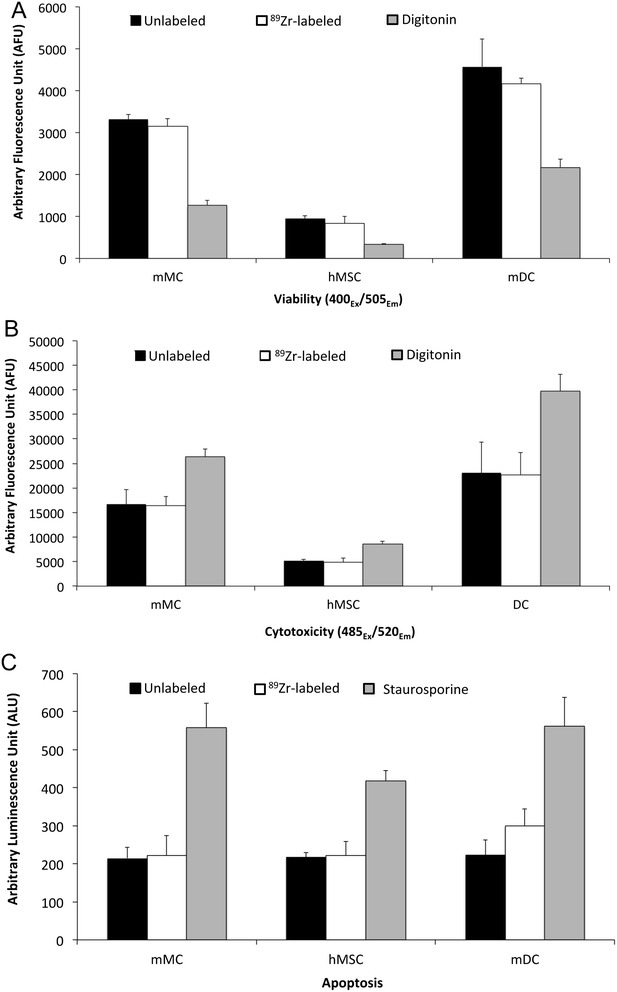
The labeling agent, (89)Zr-DBN, was prepared in 55% ± 5% decay-corrected radiochemical yield measured by silica gel iTLC. The cell labeling efficiency was 30% to 50% after 30 min labeling depending on cell type. Radioactivity concentrations of labeled cells of up to 0.5 MBq/10(6) cells were achieved without a negative effect on cellular viability. Cell efflux studies showed high stability of the radiolabel out to 7 days. Myocardially delivered (89)Zr-labeled hMSCs showed retention in the myocardium, as well as redistribution to the lung, liver, and bone. Intravenously administered (89)Zr-labeled hMSCs also distributed primarily to the lung, liver, and bone, whereas intravenous (89)Zr(HPO4)2 distributed to the liver and bone with no activity in the lung. Thus, the in vivo stability of the radiolabel on the hMSCs was evidenced.
We have developed a robust, general, and biostable (89)Zr-DBN-based cell labeling strategy with promise for wide applications of PET-based non-invasive in vivo cell trafficking.
随着近期对基于细胞的疗法和放射性标记细胞产品的兴趣增加,需要开发更强大的细胞标记和成像方法,用于体内追踪活细胞。本研究描述了一种使用正电子发射断层扫描(PET)同位素(89)Zr(半衰期T 1/2 = 78.4小时)的新型细胞标记方法的评估。(89)Zr可使PET成像测量持续数周,并利用PET成像的高灵敏度。
合成了一种新型细胞标记剂(89)Zr-去铁胺-NCS((89)Zr-DBN)。通过(89)Zr-DBN上的NCS基团与细胞表面膜蛋白上存在的伯胺基团之间的反应,将小鼠来源的黑色素瘤细胞(mMCs)、树突状细胞(mDCs)和人间充质干细胞(hMSCs)与(89)Zr-DBN共价标记。通过7天的细胞外排研究测试标记在细胞上的稳定性。通过增殖、台盼蓝和细胞毒性/凋亡测定测试标记对细胞活力的影响。通过连续PET扫描以及静脉内和心肌内注射(89)Zr标记的hMSCs后的离体生物分布,在体内小鼠模型中研究标记的稳定性。为了进行比较,在静脉注射(89)Zr磷酸氢盐((89)Zr(HPO4)2)后进行成像实验。
标记剂(89)Zr-DBN的制备放射性化学产率经硅胶iTLC测量为55%±5%(衰变校正)。根据细胞类型,标记30分钟后细胞标记效率为30%至50%。标记细胞的放射性浓度达到高达0.5 MBq/10(6)个细胞,且对细胞活力无负面影响。细胞外排研究表明放射性标记在7天内具有高稳定性。心肌递送的(89)Zr标记的hMSCs在心肌中保留,同时重新分布到肺、肝和骨。静脉内给予的(89)Zr标记的hMSCs也主要分布到肺、肝和骨,而静脉内给予的(89)Zr(HPO4)2分布到肝和骨,在肺中无活性。因此,证明了hMSCs上放射性标记的体内稳定性。
我们开发了一种强大、通用且生物稳定的基于(89)Zr-DBN的细胞标记策略,有望广泛应用于基于PET的非侵入性体内细胞运输。