Japaridze Natia, Muthuraman Muthuraman, Reinicke Christine, Moeller Friederike, Anwar Abdul Rauf, Mideksa Kidist Gebremariam, Pressler Ronit, Deuschl Günther, Stephani Ulrich, Siniatchkin Michael
Department of Neuropediatrics, Christian-Albrechts-University, Kiel, Germany.
Department of Neurology, Christian-Albrechts-University, Kiel, Germany.
PLoS One. 2015 Apr 30;10(4):e0123807. doi: 10.1371/journal.pone.0123807. eCollection 2015.
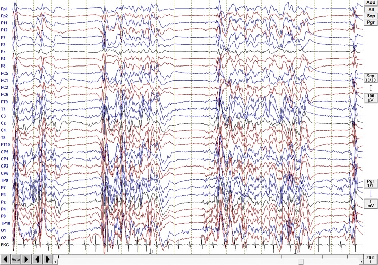
Burst-suppression (BS) is an electroencephalography (EEG) pattern consisting of alternant periods of slow waves of high amplitude (burst) and periods of so called flat EEG (suppression). It is generally associated with coma of various etiologies (hypoxia, drug-related intoxication, hypothermia, and childhood encephalopathies, but also anesthesia). Animal studies suggest that both the cortex and the thalamus are involved in the generation of BS. However, very little is known about mechanisms of BS in humans. The aim of this study was to identify the neuronal network underlying both burst and suppression phases using source reconstruction and analysis of functional and effective connectivity in EEG.
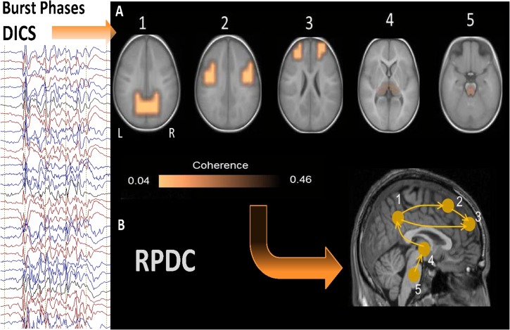
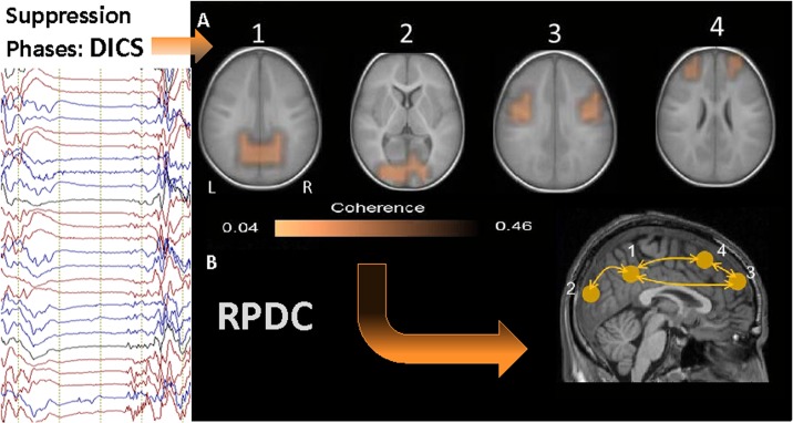
MATERIAL/METHODS: Dynamic imaging of coherent sources (DICS) was applied to EEG segments of 13 neonates and infants with burst and suppression EEG pattern. The brain area with the strongest power in the analyzed frequency (1-4 Hz) range was defined as the reference region. DICS was used to compute the coherence between this reference region and the entire brain. The renormalized partial directed coherence (RPDC) was used to describe the informational flow between the identified sources.
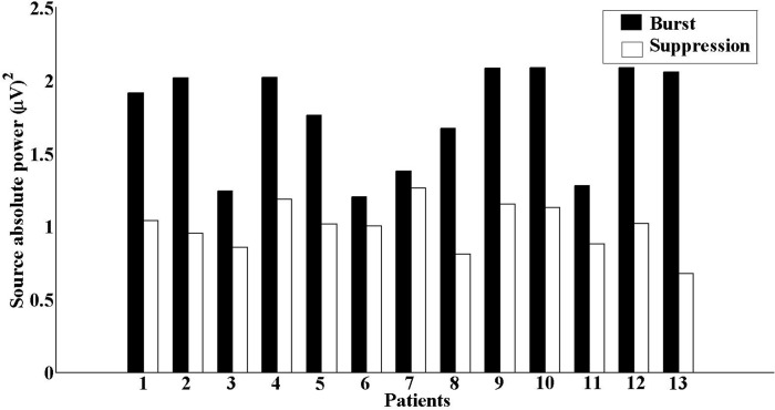
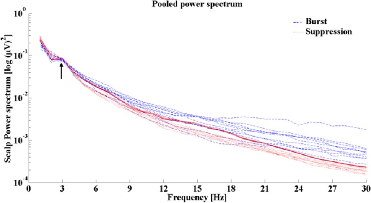
RESULTS/CONCLUSION: Delta activity during the burst phases was associated with coherent sources in the thalamus and brainstem as well as bilateral sources in cortical regions mainly frontal and parietal, whereas suppression phases were associated with coherent sources only in cortical regions. Results of the RPDC analyses showed an upwards informational flow from the brainstem towards the thalamus and from the thalamus to cortical regions, which was absent during the suppression phases. These findings may support the theory that a "cortical deafferentiation" between the cortex and sub-cortical structures exists especially in suppression phases compared to burst phases in burst suppression EEGs. Such a deafferentiation may play a role in the poor neurological outcome of children with these encephalopathies.
爆发抑制(BS)是一种脑电图(EEG)模式,由高振幅慢波的交替期(爆发)和所谓的脑电图平段期(抑制)组成。它通常与各种病因引起的昏迷相关(缺氧、药物中毒、体温过低和儿童脑病,也包括麻醉)。动物研究表明,皮层和丘脑都参与了爆发抑制的产生。然而,关于人类爆发抑制的机制知之甚少。本研究的目的是通过源重建以及脑电图功能和有效连接性分析来确定爆发期和抑制期背后的神经元网络。
材料/方法:对13名具有爆发抑制脑电图模式的新生儿和婴儿的脑电图片段应用相干源动态成像(DICS)。将分析频率(1 - 4 Hz)范围内功率最强的脑区定义为参考区域。DICS用于计算该参考区域与整个大脑之间的相干性。重新归一化的偏定向相干性(RPDC)用于描述所识别源之间的信息流。
结果/结论:爆发期的δ活动与丘脑和脑干中的相干源以及主要为额叶和顶叶的皮层区域双侧源相关,而抑制期仅与皮层区域的相干源相关。RPDC分析结果显示,存在从脑干向上流向丘脑以及从丘脑流向皮层区域的信息流,而在抑制期则不存在这种信息流。这些发现可能支持这样一种理论,即在爆发抑制脑电图中,与爆发期相比,尤其是在抑制期,皮层与皮层下结构之间存在“皮层去传入”现象。这种去传入可能在患有这些脑病儿童的不良神经学预后中起作用。