Moreira Ângela, Pereira Sofia S, Costa Madalena, Morais Tiago, Pinto Ana, Fernandes Rúben, Monteiro Mariana P
Department of Anatomy, Unit for Multidisciplinary Research in Biomedicine (UMIB), Institute for Biomedical Sciences Abel Salazar (ICBAS), University of Porto, Porto, Portugal.
Ciências Químicas e das Biomoléculas (CQB), Escola Superior de Tecnologia da Saúde do Porto do Instituto Politécnico do Porto (ESTSP-IPP), Vila Nova de Gaia, Portugal; Centro de Investigação em Saúde e Ambiente (CISA), Escola Superior de Tecnologia da Saúde do Porto do Instituto Politécnico do Porto (ESTSP-IPP), Vila Nova de Gaia, Portugal.
PLoS One. 2015 Apr 30;10(4):e0123217. doi: 10.1371/journal.pone.0123217. eCollection 2015.
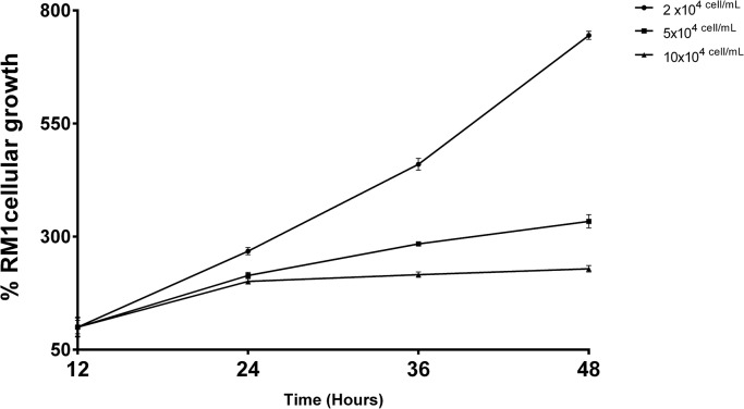
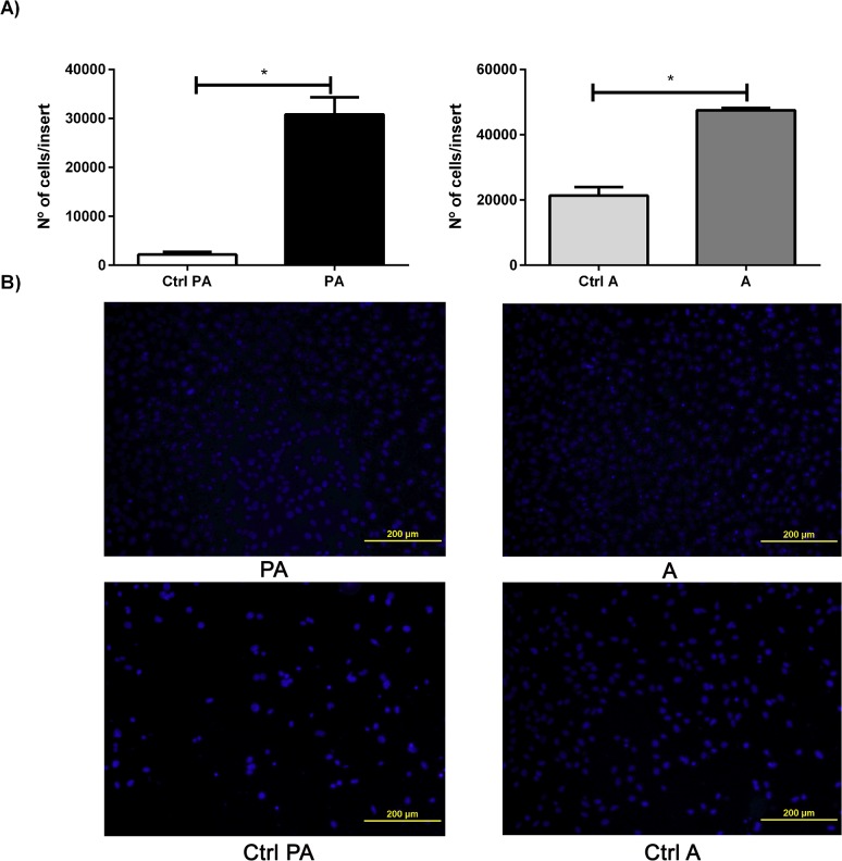
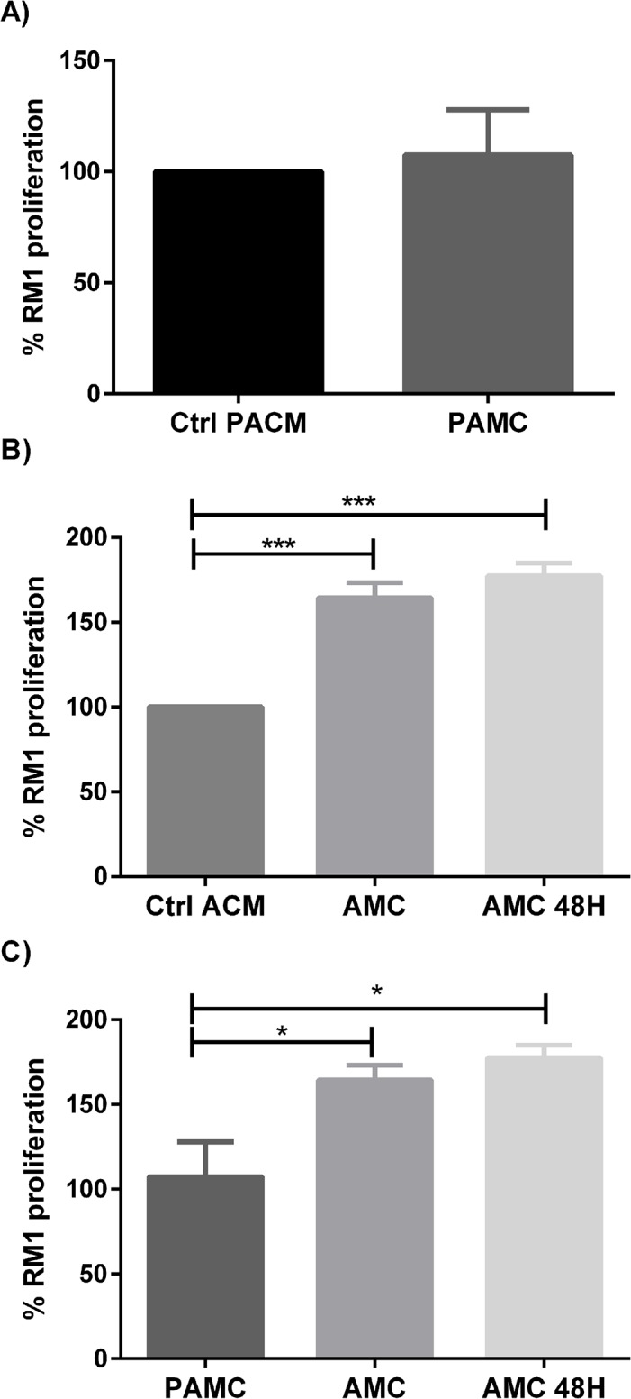
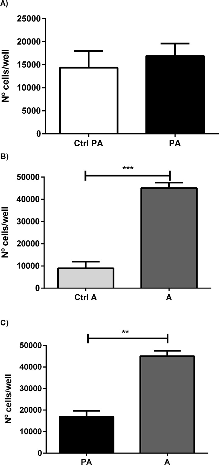
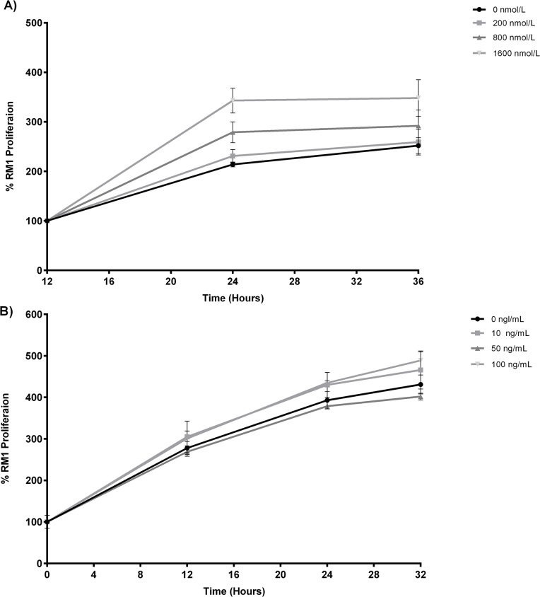
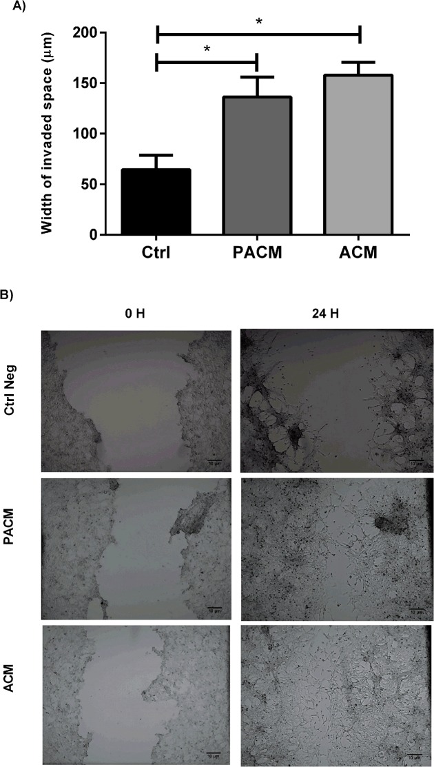
Obesity has been associated with increased incidence and risk of mortality of prostate cancer. One of the proposed mechanisms underlying this risk association is the change in adipokines expression that could promote the development and progression of the prostate tumor cells. The main goal of this study was to evaluate the effect of preadipocyte and adipocyte secretome in the proliferation, migration and invasion of androgen independent prostate carcinoma cells (RM1) and to assess cell proliferation in the presence of the adiposity signals leptin and insulin. RM1 cells were co-cultured in with preadipocytes, adipocytes or cultured in their respective conditioned medium. Cell proliferation was assessed by flow cytometry and XTT viability test. Cell migration was evaluated using a wound healing injury assay of RM1 cells cultured with conditioned media. Cellular invasion of RM1 cells co-cultured with adipocytes and preadipocytes was assessed using matrigel membranes. Preadipocyte conditioned medium was associated with a small increase in RM1 proliferation, while adipocytes conditioned media significantly increased RM1 cell proliferation (p<0.01). Adipocytes also significantly increased the RM1 cells proliferation in co-culture (p <0.01). Cell migration was higher in RM1 cells cultured with preadipocyte and adipocyte conditioned medium. RM1 cell invasion was significantly increased after co-culture with preadipocytes and adipocytes (p <0.05). Insulin also increased significantly the cell proliferation in contrast to leptin, which showed no effect. In conclusion, prostate carcinoma cells seem to be influenced by factors secreted by adipocytes that are able to increase their ability to proliferate, migrate and invade.
肥胖与前列腺癌发病率和死亡率的增加有关。这种风险关联背后提出的机制之一是脂肪因子表达的变化,这可能促进前列腺肿瘤细胞的发展和进展。本研究的主要目的是评估前脂肪细胞和脂肪细胞分泌组对雄激素非依赖性前列腺癌细胞(RM1)增殖、迁移和侵袭的影响,并评估在肥胖信号瘦素和胰岛素存在下的细胞增殖。RM1细胞与前脂肪细胞、脂肪细胞共培养或在其各自的条件培养基中培养。通过流式细胞术和XTT活力试验评估细胞增殖。使用与条件培养基一起培养的RM1细胞的伤口愈合损伤试验评估细胞迁移。使用基质胶膜评估与脂肪细胞和前脂肪细胞共培养的RM1细胞的细胞侵袭。前脂肪细胞条件培养基与RM1增殖的小幅增加有关,而脂肪细胞条件培养基显著增加RM1细胞增殖(p<0.01)。脂肪细胞在共培养中也显著增加RM1细胞增殖(p<0.01)。用前脂肪细胞和脂肪细胞条件培养基培养的RM1细胞的细胞迁移更高。与前脂肪细胞和脂肪细胞共培养后,RM1细胞侵袭显著增加(p<0.05)。与瘦素相比,胰岛素也显著增加细胞增殖,瘦素无作用。总之,前列腺癌细胞似乎受到脂肪细胞分泌的因子的影响,这些因子能够增加其增殖、迁移和侵袭能力。