Narematsu Mayu, Kamimura Tatsuya, Yamagishi Toshiyuki, Fukui Mitsuru, Nakajima Yuji
Department of Anatomy and Cell Biology, Graduate School of Medicine, Osaka City University, Osaka, Japan (M.N., T.K., T.Y., Y.N.).
Laboratory of Statics, Graduate School of Medicine, Osaka City University, Osaka, Japan (M.F.).
J Am Heart Assoc. 2015 Apr 30;4(5):e001889. doi: 10.1161/JAHA.115.001889.
Transposition of the great arteries is one of the most commonly diagnosed conotruncal heart defects at birth, but its etiology is largely unknown. The anterior heart field (AHF) that resides in the anterior pharyngeal arches contributes to conotruncal development, during which heart progenitors that originated from the left and right AHF migrate to form distinct conotruncal regions. The aim of this study is to identify abnormal AHF development that causes the morphology of transposition of the great arteries.
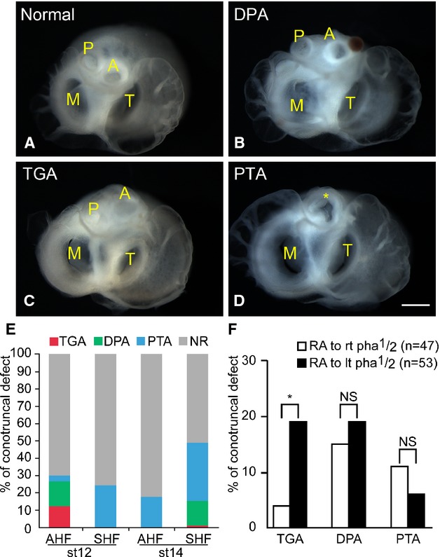
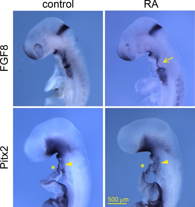
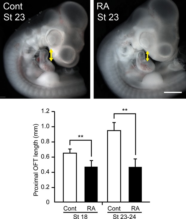
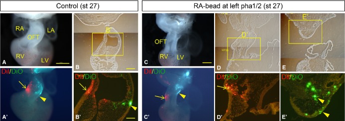
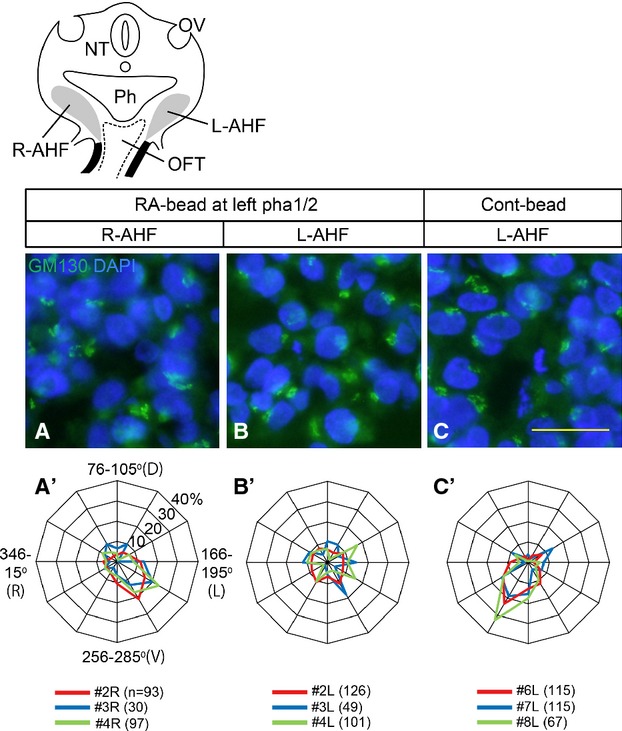
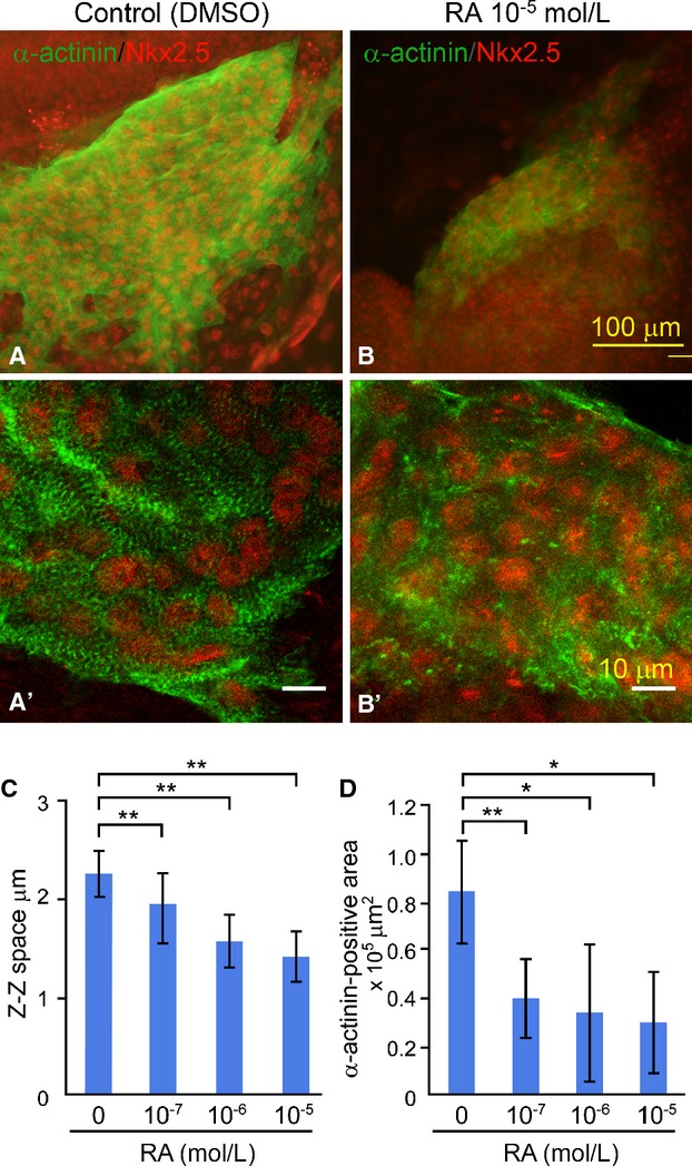
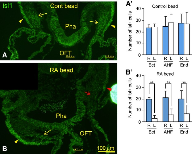
We placed a retinoic acid-soaked bead on the left or the right or on both sides of the AHF of stage 12 to 14 chick embryos and examined the conotruncal heart defect at stage 34. Transposition of the great arteries was diagnosed at high incidence in embryos for which a retinoic acid-soaked bead had been placed in the left AHF at stage 12. Fluorescent dye tracing showed that AHF exposed to retinoic acid failed to contribute to conotruncus development. FGF8 and Isl1 expression were downregulated in retinoic acid-exposed AHF, and differentiation and expansion of cardiomyocytes were suppressed in cultured AHF in medium supplemented with retinoic acid.
The left AHF at the early looped heart stage, corresponding to Carnegie stages 10 to 11 (28 to 29 days after fertilization) in human embryos, is the region of the impediment that causes the morphology of transposition of the great arteries.
大动脉转位是出生时最常被诊断出的圆锥动脉干心脏缺陷之一,但其病因在很大程度上尚不清楚。位于咽前弓的前心脏原基有助于圆锥动脉干的发育,在此过程中,源自左、右前心脏原基的心脏祖细胞迁移以形成不同的圆锥动脉干区域。本研究的目的是确定导致大动脉转位形态的前心脏原基异常发育情况。
我们将浸有视黄酸的珠子放置在第12至14期鸡胚前心脏原基的左侧、右侧或两侧,并在第34期检查圆锥动脉干心脏缺陷情况。在第12期将浸有视黄酸的珠子置于左前心脏原基的胚胎中,大动脉转位的发生率很高。荧光染料追踪显示,暴露于视黄酸的前心脏原基未能促进圆锥动脉干的发育。在暴露于视黄酸的前心脏原基中,FGF8和Isl1表达下调,并且在添加视黄酸的培养基中培养的前心脏原基中,心肌细胞的分化和扩增受到抑制。
在早期心脏成环阶段的左前心脏原基,相当于人类胚胎的卡内基阶段10至11期(受精后28至29天),是导致大动脉转位形态的阻碍区域。