Li Pengfei, Guo Youming, Bledsoe Grant, Yang Zhi-Rong, Fan Hongkuan, Chao Lee, Chao Julie
Department of Biochemistry and Molecular Biology, Medical University of South Carolina, 173 Ashley Ave, Charleston, SC, 29425-2211, USA.
Department of Neurosciences, Medical University of South Carolina, 173 Ashley Ave, Charleston, SC, 29425-2211, USA.
Crit Care. 2015 May 1;19(1):200. doi: 10.1186/s13054-015-0919-4.
Kallistatin levels in the circulation are reduced in patients with sepsis and liver disease. Transgenic mice expressing kallistatin are resistant to lipopolysaccharide (LPS)-induced mortality. Here, we investigated the effect of kallistatin on survival and organ damage in mouse models of established sepsis.
Mice were rendered septic by cecal ligation and puncture (CLP), or endotoxemic by LPS injection. Recombinant human kallistatin was administered intravenously six hours after CLP, or intraperitoneally four hours after LPS challenge. The effect of kallistatin treatment on organ damage was examined one day after sepsis initiation, and mouse survival was monitored for four to six days.
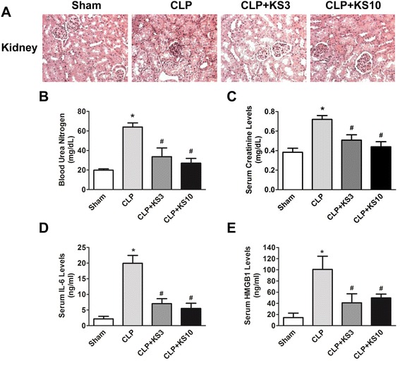
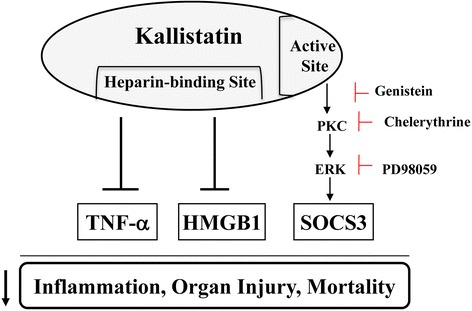
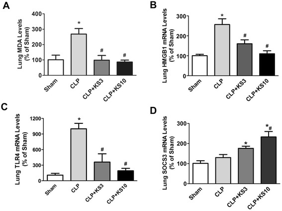
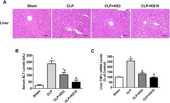
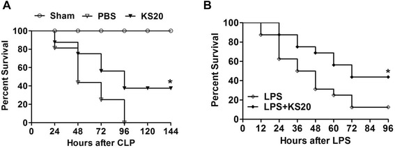
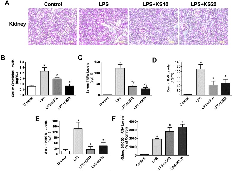
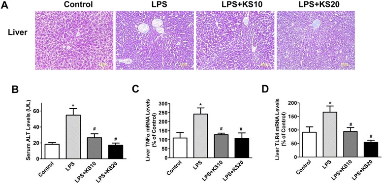
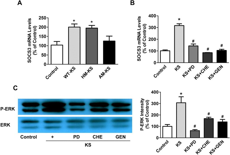
Human kallistatin was detected in mouse serum of kallistatin-treated mice. Kallistatin significantly reduced CLP-induced renal injury as well as blood urea nitrogen, serum creatinine, interleukin-6 (IL-6), and high mobility group box-1 (HMGB1) levels. In the lung, kallistatin decreased malondialdehyde levels and HMGB1 and toll-like receptor-4 (TLR4) synthesis, but increased suppressor of cytokine signaling-3 (SOCS3) expression. Moreover, kallistatin attenuated liver injury, serum alanine transaminase (ALT) levels and hepatic tumor necrosis factor-α (TNF-α) synthesis. Furthermore, delayed kallistatin administration improved survival in CLP mice by 38%, and LPS-treated mice by 42%. In LPS-induced endotoxemic mice, kallistatin attenuated kidney damage in association with reduced serum creatinine, IL-6 and HMGB1 levels, and increased renal SOCS3 expression. Kallistatin also decreased liver injury in conjunction with diminished serum ALT levels and hepatic TNF-α and TLR4 expression. In cultured macrophages, kallistatin through its active site increased SOCS3 expression, but this effect was blocked by inhibitors of tyrosine kinase, protein kinase C and extracellular signal-regulated kinase (ERK), indicating that kallistatin stimulates a tyrosine-kinase-protein kinase C-ERK signaling pathway.
This is the first study to demonstrate that delayed human kallistatin administration is effective in attenuating multi-organ injury, inflammation and mortality in mouse models of polymicrobial infection and endotoxemia. Thus, kallistatin therapy may provide a promising approach for the treatment of sepsis in humans.
脓毒症和肝病患者循环中的抑癌素M水平降低。表达抑癌素M的转基因小鼠对脂多糖(LPS)诱导的死亡具有抗性。在此,我们研究了抑癌素M对已建立的脓毒症小鼠模型中生存和器官损伤的影响。
通过盲肠结扎和穿刺(CLP)使小鼠发生脓毒症,或通过注射LPS使其发生内毒素血症。在CLP后6小时静脉注射重组人抑癌素M,或在LPS攻击后4小时腹腔注射。在脓毒症开始后1天检查抑癌素M治疗对器官损伤的影响,并监测小鼠存活4至6天。
在接受抑癌素M治疗的小鼠血清中检测到人类抑癌素M。抑癌素M显著减轻CLP诱导的肾损伤以及血尿素氮、血清肌酐、白细胞介素-6(IL-6)和高迁移率族蛋白B1(HMGB1)水平。在肺中,抑癌素M降低丙二醛水平以及HMGB1和Toll样受体4(TLR4)的合成,但增加细胞因子信号转导抑制因子3(SOCS3)的表达。此外,抑癌素M减轻肝损伤、血清丙氨酸转氨酶(ALT)水平和肝肿瘤坏死因子-α(TNF-α)的合成。此外,延迟给予抑癌素M可使CLP小鼠的存活率提高38%,使LPS处理的小鼠存活率提高42%。在LPS诱导的内毒素血症小鼠中,抑癌素M减轻肾损伤,同时降低血清肌酐、IL-6和HMGB1水平,并增加肾SOCS3表达。抑癌素M还减轻肝损伤,同时降低血清ALT水平以及肝TNF-α和TLR4表达。在培养的巨噬细胞中,抑癌素M通过其活性位点增加SOCS3表达,但这种作用被酪氨酸激酶、蛋白激酶C和细胞外信号调节激酶(ERK)抑制剂阻断,表明抑癌素M刺激酪氨酸激酶-蛋白激酶C-ERK信号通路。
这是第一项证明延迟给予人类抑癌素M可有效减轻多微生物感染和内毒素血症小鼠模型中的多器官损伤、炎症和死亡率的研究。因此,抑癌素M治疗可能为人类脓毒症的治疗提供一种有前景的方法。