Li Yue, Kang Guobin, Duan Lijie, Lu Wuxun, Katze Michael G, Lewis Mark G, Haase Ashley T, Li Qingsheng
College of Life Sciences, Nankai University, Tianjin, People's Republic of China; Nebraska Center for Virology, School of Biological Sciences, University of Nebraska-Lincoln, Lincoln, Nebraska, United States of America.
Nebraska Center for Virology, School of Biological Sciences, University of Nebraska-Lincoln, Lincoln, Nebraska, United States of America.
PLoS One. 2015 May 1;10(5):e0125500. doi: 10.1371/journal.pone.0125500. eCollection 2015.
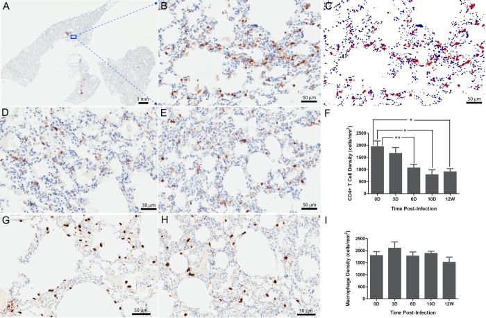
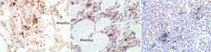
HIV-1 depletes CD4+ T cells in the blood, lymphatic tissues, gut and lungs. Here we investigated the relationship between depletion and infection of CD4+ T cells in the lung parenchyma. The lungs of 38 Indian rhesus macaques in early to later stages of SIVmac251 infection were examined, and the numbers of CD4+ T cells and macrophages plus the frequency of SIV RNA+ cells were quantified. We showed that SIV infected macrophages in the lung parenchyma, but only in small numbers except in the setting of interstitial inflammation where large numbers of SIV RNA+ macrophages were detected. However, even in this setting, the number of macrophages was not decreased. By contrast, there were few infected CD4+ T cells in lung parenchyma, but CD4+ T cells were nonetheless depleted by unknown mechanisms. The CD4+ T cells in lung parenchyma were depleted even though they were not productively infected, whereas SIV can infect large numbers of macrophages in the setting of interstitial inflammation without depleting them. These observations point to the need for future investigations into mechanisms of CD4+ T cell depletion at this mucosal site, and into mechanisms by which macrophage populations are maintained despite high levels of infection. The large numbers of SIV RNA+ macrophages in lungs in the setting of interstitial inflammation indicates that lung macrophages can be an important source for SIV persistent infection.
HIV-1会消耗血液、淋巴组织、肠道和肺部中的CD4+ T细胞。在此,我们研究了肺实质中CD4+ T细胞的消耗与感染之间的关系。对38只处于SIVmac251感染早期至晚期的印度恒河猴的肺部进行了检查,并对CD4+ T细胞和巨噬细胞的数量以及SIV RNA+细胞的频率进行了定量分析。我们发现,SIV感染了肺实质中的巨噬细胞,但数量很少,除了在间质性炎症情况下会检测到大量SIV RNA+巨噬细胞。然而,即使在这种情况下,巨噬细胞的数量也没有减少。相比之下,肺实质中被感染的CD4+ T细胞很少,但CD4+ T细胞却通过未知机制被消耗。肺实质中的CD4+ T细胞即使没有被有效感染也会被消耗,而SIV在间质性炎症情况下可以感染大量巨噬细胞却不会使其减少。这些观察结果表明,未来需要研究该黏膜部位CD4+ T细胞消耗的机制,以及巨噬细胞群体在高感染水平下仍能维持的机制。间质性炎症情况下肺部大量的SIV RNA+巨噬细胞表明,肺巨噬细胞可能是SIV持续感染的重要来源。