Almeida Etanaiara, Mascarenhas Bruno Araújo, Cerqueira Arlei, Medrado Alena Ribeiro Alves Peixoto
Department of Basic Science and Bahian School of Medicine and Public Health, Salvador, Brazil.
Department of Diagnosis and Therapeutics, Bahia Federal University, Salvador, Bahia, Brazil.
J Oral Maxillofac Pathol. 2014 Sep-Dec;18(3):464-8. doi: 10.4103/0973-029X.151357.
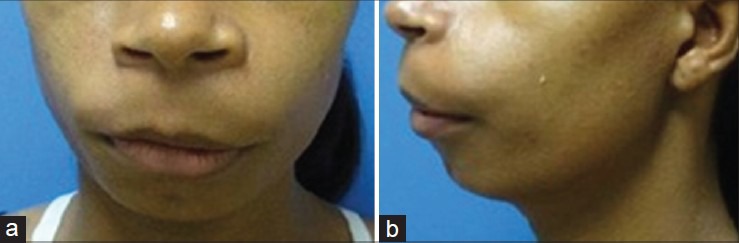
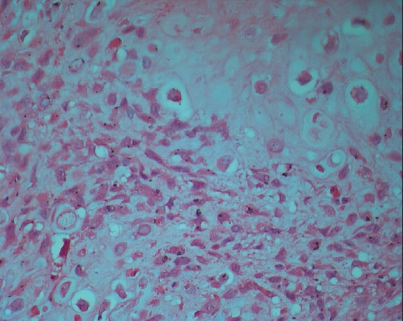
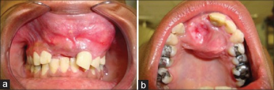
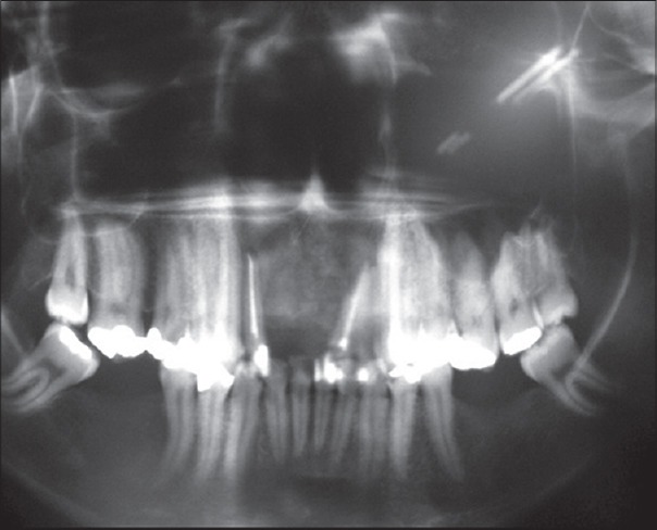
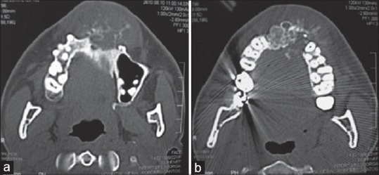
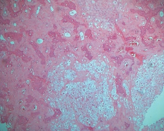
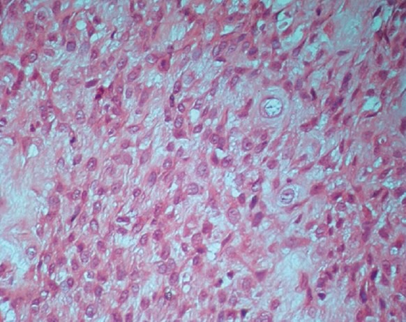
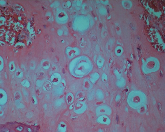
The purpose of this paper is to report a case of chondroblastic osteosarcoma in the region of the maxilla, with 5 months of evolution. The term osteosarcoma refers to a heterogeneous group of malignancies with bone formation or mesenchymal tissue with histopathological evidence of osteogenic differentiation. The pattern of chondroblastic osteosarcoma represents 25% of all reported cases of this neoplasm. Its histopathological diagnosis is based on the predominance of a chondroid matrix formed in the midst of neoplastic cells. A woman patient, 27-year old, melanoderm, presented on extraoral exam with facial asymmetry caused by the a swelling in the premaxillary region with upper lip protrusion. Intraoral exam showed a maxillary tumefaction with involvement of the vestibular and palatine regions. The computerized tomography (CT) analysis exhibited a radiolucent mass with dispersed areas of radiopacity, with poorly defined and indistinct peripheral edges. The patient was subjected to incisional biopsy and histopathological examination showed the presence of a malignant neoplasm of mesenchymal origin characterized by the presence of irregular bone trabeculae dispersed among mildly atypical chondroblastic cells. The World Health Organization (WHO) recognizes several variants that differ in location, clinical behavior and degree of cellular atypia. The conventional or classical osteosarcoma is the most frequent variant, which develops within the medullary bone. This report illustrates the rapid evolution of one of the histological variants of osteosarcoma.
本文旨在报告一例上颌骨区域的软骨母细胞性骨肉瘤病例,病程为5个月。骨肉瘤一词指的是一组异质性恶性肿瘤,具有骨形成或间充质组织,并有成骨分化的组织病理学证据。软骨母细胞性骨肉瘤模式占该肿瘤所有报告病例的25%。其组织病理学诊断基于肿瘤细胞中形成的软骨样基质占优势。一名27岁的黑皮肤女性患者,口外检查显示前上颌区域肿胀伴上唇突出,导致面部不对称。口内检查显示上颌肿物累及前庭和腭部区域。计算机断层扫描(CT)分析显示一个透射线肿物,有散在的不透射线区域,周边边缘模糊不清。患者接受了切开活检,组织病理学检查显示存在间叶源性恶性肿瘤,其特征为不规则骨小梁散在于轻度非典型软骨母细胞之间。世界卫生组织(WHO)认可几种在位置、临床行为和细胞异型程度上有所不同的变体。传统或经典骨肉瘤是最常见的变体,发生于髓腔内骨。本报告说明了骨肉瘤的一种组织学变体的快速发展情况。