Rojanaporn Duangnate, Kaliki Swathi, Ferenczy Sandor R, Shields Carol L
Department of Ophthalmology, Ocular Oncology Service, Wills Eye Hospital, Thomas Jefferson University, Philadelphia, PA, USA ; Department of Ophthalmology, Faculty of Medicine, Ramathibodi Hospital, Mahidol University, Bangkok, Thailand.
Department of Ophthalmology, Ocular Oncology Service, Wills Eye Hospital, Thomas Jefferson University, Philadelphia, PA, USA ; Ocular Oncology Service, L.V. Prasad Eye Institute, Hyderabad, India.
Middle East Afr J Ophthalmol. 2015 Apr-Jun;22(2):192-7. doi: 10.4103/0974-9233.150629.
The purpose was to evaluate the features of circumscribed choroidal hemangioma using spectral-domain enhanced depth imaging optical coherence tomography (EDI-OCT).
Retrospective observational case series.
Ten patients with newly diagnosed circumscribed choroidal hemangioma.
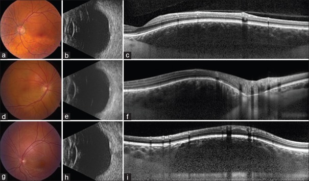
Spectral-domain EDI-OCT was performed with a Heidelberg Spectralis HRA + OCT (Heidelberg Engineering, Heidelberg, Germany).
Tumor thickness and EDI-OCT features.
The mean tumor diameter for all eyes was 5.4 mm and mean tumor thickness was 1187 μm by EDI-OCT compared to 2400 μm by ultrasonography. EDI-OCT imaged all tumors as smooth with a gently sloping anterior contour, gradual choroidal expansion, expansion of medium and large size choroidal vessels without compression of choriocapillaris, and intact Bruch's membrane (n = 10, 100%). The height of the medium and large choroidal vessels within the tumor compared to normal medium and large vessels was comparatively increased by a mean of 265% (medium vessels) and 576% (large vessels). Outer retinal abnormalities included subretinal fluid (n = 7, 70%), lipofuscin deposition (n = 1, 10%), irregularity and thinning of retinal pigment epithelium and absence or irregularity of the ellipsoid layer (n = 4, 40%), absent external limiting membrane (n = 2, 20%), and disruption of the outer nuclear layer and outer plexiform layer (n = 3, 30%). The inner retinal abnormalities included irregularity of inner nuclear layer and structural loss or edema of inner plexiform layer (n = 3, 30%). The ganglion cell layer and nerve fiber layer were intact (n = 10, 100%).
EDI-OCT of circumscribed choroidal hemangiomas depicts a smooth, gently sloping choroidal mass with expansion of medium and large size choroidal vessels without compression of the choriocapillaris. Structural abnormalities of outer and inner retinal layers were noted.
使用光谱域增强深度成像光学相干断层扫描(EDI-OCT)评估局限性脉络膜血管瘤的特征。
回顾性观察病例系列。
10例新诊断的局限性脉络膜血管瘤患者。
使用德国海德堡海德堡工程公司的海德堡Spectralis HRA + OCT进行光谱域EDI-OCT检查。
肿瘤厚度和EDI-OCT特征。
所有患眼的平均肿瘤直径为5.4 mm,通过EDI-OCT测量的平均肿瘤厚度为1187μm,而超声检查测得的平均肿瘤厚度为2400μm。EDI-OCT显示所有肿瘤表面光滑,前部轮廓呈缓坡状,脉络膜逐渐扩张,中大型脉络膜血管扩张而无脉络膜毛细血管受压,且布鲁赫膜完整(n = 10,100%)。肿瘤内中大型脉络膜血管相对于正常中大型血管的高度平均分别增加了265%(中型血管)和576%(大型血管)。视网膜外层异常包括视网膜下液(n = 7,70%)、脂褐素沉积(n = 1,10%)、视网膜色素上皮不规则和变薄以及椭圆体层缺失或不规则(n = 4,40%)、外界膜缺失(n = 2,20%),以及外核层和外丛状层破坏(n = 3,30%)。视网膜内层异常包括内核层不规则以及内丛状层结构缺失或水肿(n = 3,30%)。神经节细胞层和神经纤维层完整(n = 10,100%)。
局限性脉络膜血管瘤的EDI-OCT表现为表面光滑、前部呈缓坡状的脉络膜肿物,伴有中大型脉络膜血管扩张且无脉络膜毛细血管受压。视网膜外层和内层均存在结构异常。