• 文献检索
  • 文档翻译
  • 深度研究
  • 学术资讯
  • Suppr Zotero 插件Zotero 插件
  • 邀请有礼
  • 套餐&价格
  • 历史记录
应用&插件
Suppr Zotero 插件Zotero 插件浏览器插件Mac 客户端Windows 客户端微信小程序
定价
高级版会员购买积分包购买API积分包
服务
文献检索文档翻译深度研究API 文档MCP 服务
关于我们
关于 Suppr公司介绍联系我们用户协议隐私条款
关注我们

Suppr 超能文献

核心技术专利:CN118964589B侵权必究
粤ICP备2023148730 号-1Suppr @ 2026

文献检索

告别复杂PubMed语法,用中文像聊天一样搜索,搜遍4000万医学文献。AI智能推荐,让科研检索更轻松。

立即免费搜索

文件翻译

保留排版,准确专业,支持PDF/Word/PPT等文件格式,支持 12+语言互译。

免费翻译文档

深度研究

AI帮你快速写综述,25分钟生成高质量综述,智能提取关键信息,辅助科研写作。

立即免费体验

视网膜和脉络膜肿瘤的光学相干断层扫描

Optical coherence tomography of retinal and choroidal tumors.

作者信息

Say Emil Anthony T, Shah Sanket U, Ferenczy Sandor, Shields Carol L

机构信息

Oncology Service, Wills Eye Institute, Thomas Jefferson University, Suite 1440, 840 Walnut Street, Philadelphia, PA 19107, USA.

出版信息

J Ophthalmol. 2012;2012:385058. doi: 10.1155/2011/385058. Epub 2011 Jun 8.

DOI:10.1155/2011/385058
PMID:23008756
原文链接:https://pmc.ncbi.nlm.nih.gov/articles/PMC3139893/
Abstract

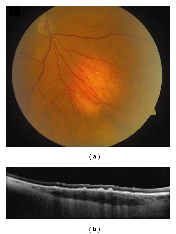
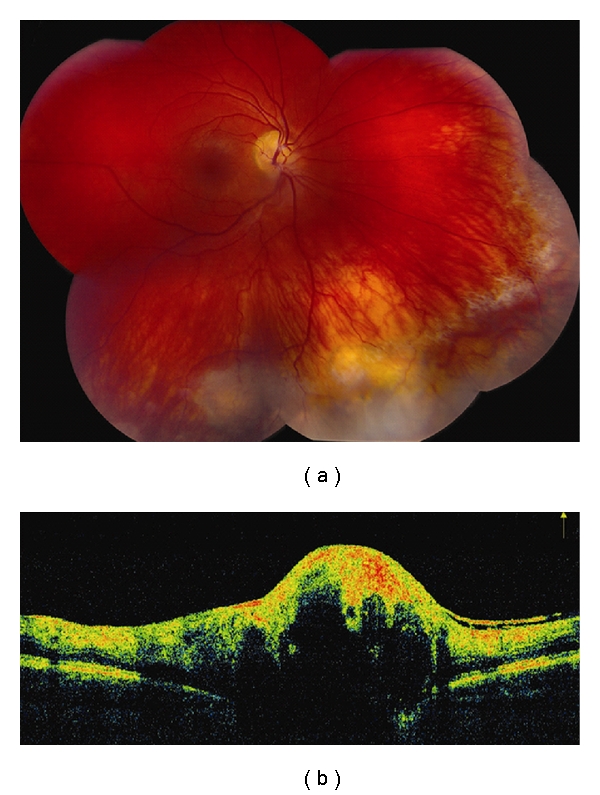
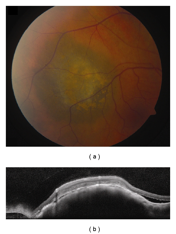
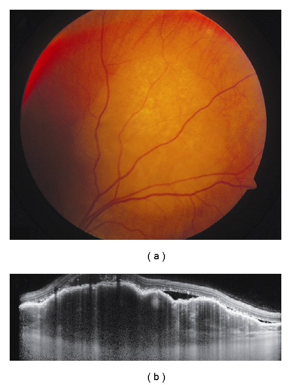
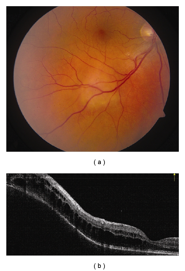
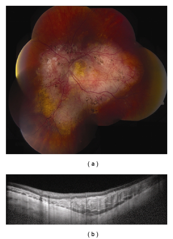
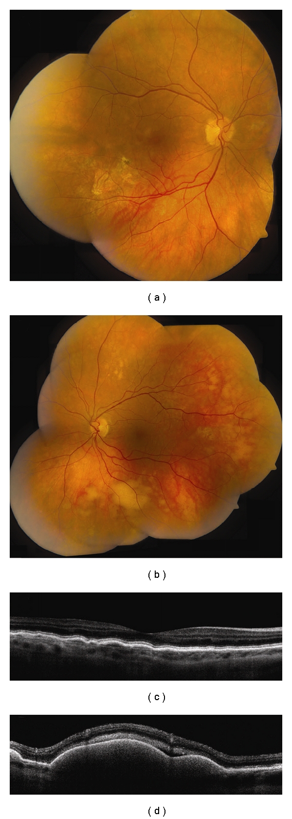
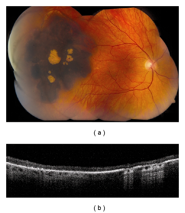
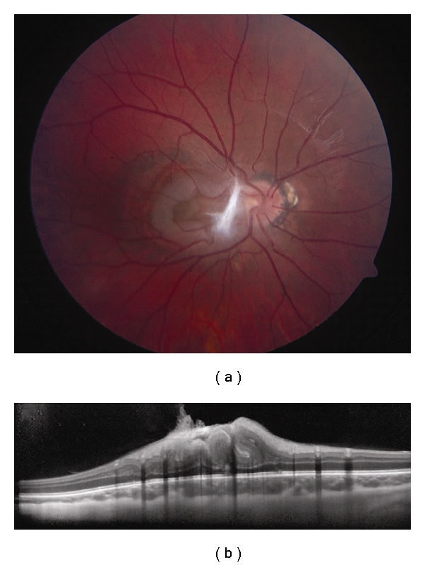
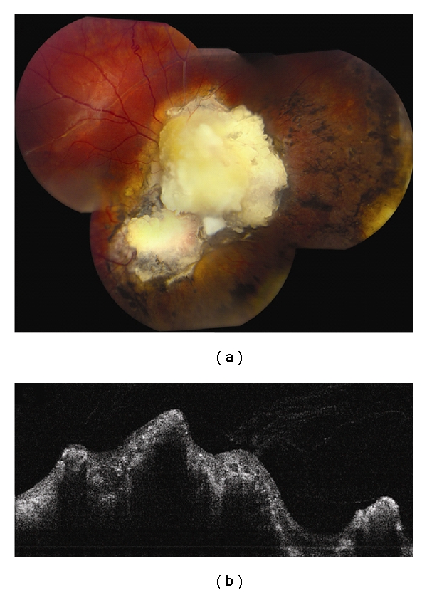
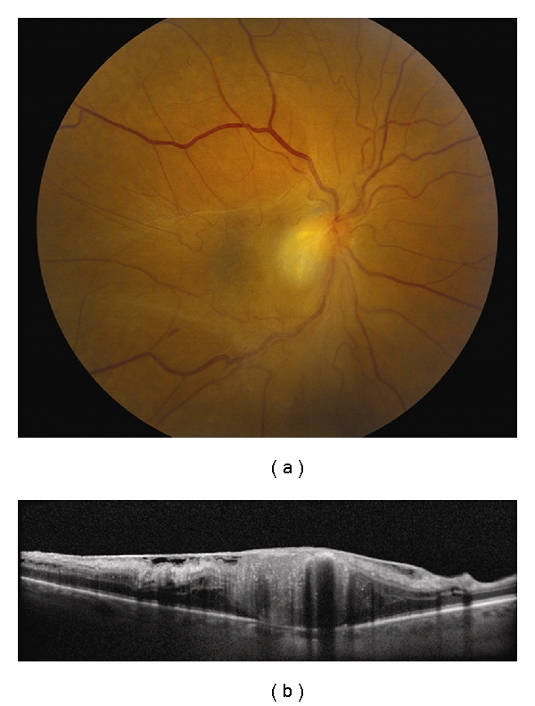
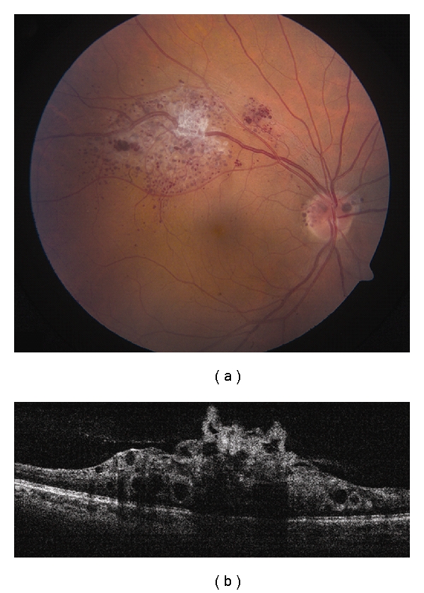
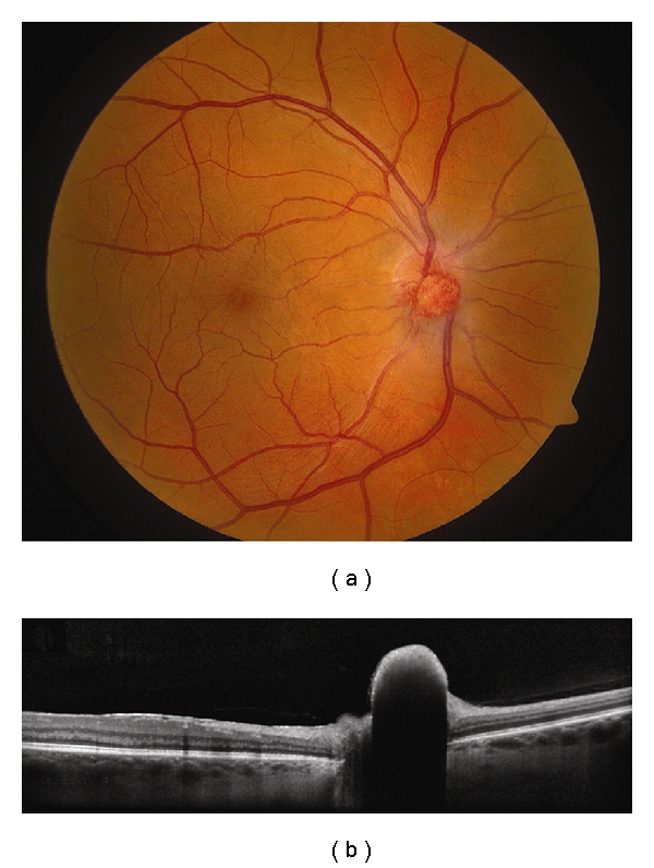
Optical coherence tomography (OCT) has revolutionized the field of ophthalmology since its introduction 20 years ago. Originally intended primarily for retina specialists to image the macula, it has found its role in other subspecialties that include glaucoma, cornea, and ocular oncology. In ocular oncology, OCT provides axial resolution to approximately 7 microns with cross-sectional images of the retina, delivering valuable information on the effects of intraocular tumors on the retinal architecture. Some effects include retinal edema, subretinal fluid, retinal atrophy, photoreceptor loss, outer retinal thinning, and retinal pigment epithelial detachment. With more advanced technology, OCT now provides imaging deeper into the choroid using a technique called enhanced depth imaging. This allows characterization of the thickness and reflective quality of small (<3 mm thick) choroidal lesions including choroidal nevus and melanoma. Future improvements in image resolution and depth will allow better understanding of the mechanisms of visual loss, tumor growth, and tumor management.

摘要

自20年前引入以来,光学相干断层扫描(OCT)彻底改变了眼科领域。它最初主要供视网膜专家用于黄斑成像,如今已在青光眼、角膜和眼部肿瘤学等其他亚专业领域发挥作用。在眼部肿瘤学中,OCT能提供约7微米的轴向分辨率,生成视网膜的横截面图像,提供有关眼内肿瘤对视网膜结构影响的有价值信息。一些影响包括视网膜水肿、视网膜下液、视网膜萎缩、光感受器丧失、外层视网膜变薄以及视网膜色素上皮脱离。随着技术的不断进步,OCT现在使用一种称为增强深度成像的技术,能够对脉络膜进行更深层次的成像。这使得能够对小的(厚度小于3毫米)脉络膜病变(包括脉络膜痣和黑色素瘤)的厚度和反射特性进行表征。图像分辨率和深度的未来改进将有助于更好地理解视力丧失机制、肿瘤生长和肿瘤管理。

https://cdn.ncbi.nlm.nih.gov/pmc/blobs/9d55/3139893/88cee099b9b7/JOP2012-385058.013.jpg
https://cdn.ncbi.nlm.nih.gov/pmc/blobs/9d55/3139893/482f72ba47de/JOP2012-385058.001.jpg
https://cdn.ncbi.nlm.nih.gov/pmc/blobs/9d55/3139893/de32e6746a09/JOP2012-385058.002.jpg
https://cdn.ncbi.nlm.nih.gov/pmc/blobs/9d55/3139893/48db405b1122/JOP2012-385058.003.jpg
https://cdn.ncbi.nlm.nih.gov/pmc/blobs/9d55/3139893/aa729d788964/JOP2012-385058.004.jpg
https://cdn.ncbi.nlm.nih.gov/pmc/blobs/9d55/3139893/4ab0029943a2/JOP2012-385058.005.jpg
https://cdn.ncbi.nlm.nih.gov/pmc/blobs/9d55/3139893/2f8248a31b81/JOP2012-385058.006.jpg
https://cdn.ncbi.nlm.nih.gov/pmc/blobs/9d55/3139893/2c5313e1eb72/JOP2012-385058.007.jpg
https://cdn.ncbi.nlm.nih.gov/pmc/blobs/9d55/3139893/32f098fa3231/JOP2012-385058.008.jpg
https://cdn.ncbi.nlm.nih.gov/pmc/blobs/9d55/3139893/40756b6be455/JOP2012-385058.009.jpg
https://cdn.ncbi.nlm.nih.gov/pmc/blobs/9d55/3139893/200f08ad25e4/JOP2012-385058.010.jpg
https://cdn.ncbi.nlm.nih.gov/pmc/blobs/9d55/3139893/7d8d95595710/JOP2012-385058.011.jpg
https://cdn.ncbi.nlm.nih.gov/pmc/blobs/9d55/3139893/8d196a961873/JOP2012-385058.012.jpg
https://cdn.ncbi.nlm.nih.gov/pmc/blobs/9d55/3139893/88cee099b9b7/JOP2012-385058.013.jpg

相似文献

1
Optical coherence tomography of retinal and choroidal tumors.视网膜和脉络膜肿瘤的光学相干断层扫描
J Ophthalmol. 2012;2012:385058. doi: 10.1155/2011/385058. Epub 2011 Jun 8.
2
Optical coherence tomography of retinal and choroidal tumors.视网膜和脉络膜肿瘤的光学相干断层扫描
J Ophthalmol. 2011;2011:385058. doi: 10.1155/2011/385058. Epub 2011 Jul 18.
3
Enhanced depth imaging optical coherence tomography of small choroidal melanoma: comparison with choroidal nevus.小脉络膜黑色素瘤的增强深度成像光学相干断层扫描:与脉络膜痣的比较
Arch Ophthalmol. 2012 Jul;130(7):850-6. doi: 10.1001/archophthalmol.2012.1135.
4
Enhanced depth imaging optical coherence tomography of intraocular tumors: from placid to seasick to rock and rolling topography--the 2013 Francesco Orzalesi Lecture.眼内肿瘤的增强深度成像光学相干断层扫描:从平静到晕船再到起伏地形——2013年弗朗切斯科·奥尔扎莱西讲座
Retina. 2014 Aug;34(8):1495-512. doi: 10.1097/IAE.0000000000000288.
5
Optical coherence tomography of choroidal nevus in 120 patients.120例脉络膜痣的光学相干断层扫描
Retina. 2005 Apr-May;25(3):243-52. doi: 10.1097/00006982-200504000-00001.
6
Review of optical coherence tomography for intraocular tumors.用于眼内肿瘤的光学相干断层扫描综述。
Curr Opin Ophthalmol. 2005 Jun;16(3):141-54. doi: 10.1097/01.icu.0000158258.01681.40.
7
[Pathophysiology of macular diseases--morphology and function].黄斑疾病的病理生理学——形态与功能
Nippon Ganka Gakkai Zasshi. 2011 Mar;115(3):238-74; discussion 275.
8
3D Spectral domain optical coherence tomography findings in choroidal tumors.脉络膜肿瘤的三维光谱域光学相干断层扫描结果
Eur J Ophthalmol. 2011 May-Jun;21(3):271-5. doi: 10.5301/EJO.2010.5848.
9
Limitations of imaging choroidal tumors in vivo by optical coherence tomography.光学相干断层扫描对脉络膜肿瘤进行体内成像的局限性。
Graefes Arch Clin Exp Ophthalmol. 1998 Aug;236(8):588-92. doi: 10.1007/s004170050126.
10
Enhanced depth imaging optical coherence tomography of choroidal nevus in 104 cases.104 例脉络膜黑色素瘤的增强深度成像光相干断层扫描。
Ophthalmology. 2012 May;119(5):1066-72. doi: 10.1016/j.ophtha.2011.11.001. Epub 2012 Jan 31.

引用本文的文献

1
The Small Choroidal Melanoma: Diagnosis, Treatment Outcomes, and Unending Controversy.小脉络膜黑色素瘤:诊断、治疗结果及无尽的争议
Semin Ophthalmol. 2025 Mar 21:1-10. doi: 10.1080/08820538.2025.2480188.
2
Trends in Research Related to Ophthalmic OCT Imaging From 2011 to 2020: A Bibliometric Analysis.2011年至2020年眼科光学相干断层扫描成像相关研究趋势:文献计量分析
Front Med (Lausanne). 2022 Apr 27;9:820706. doi: 10.3389/fmed.2022.820706. eCollection 2022.
3
Interocular asymmetry of choroidal thickness and vascularity index measurements in normal eyes assessed by swept-source optical coherence tomography.

本文引用的文献

1
Optical coherence tomography enhanced depth imaging of choroidal tumors.光学相干断层扫描增强深度成像在脉络膜肿瘤中的应用。
Am J Ophthalmol. 2011 Apr;151(4):586-593.e2. doi: 10.1016/j.ajo.2010.09.028. Epub 2011 Jan 22.
2
Current update on retinoblastoma.视网膜母细胞瘤的最新进展
Int Ophthalmol Clin. 2011 Winter;51(1):77-91. doi: 10.1097/IIO.0b013e3182010f29.
3
3D Spectral domain optical coherence tomography findings in choroidal tumors.脉络膜肿瘤的三维光谱域光学相干断层扫描结果
通过扫频光学相干断层扫描评估正常眼脉络膜厚度和血管指数测量的眼间不对称性。
Quant Imaging Med Surg. 2022 Jan;12(1):781-795. doi: 10.21037/qims-21-813.
4
Hidden Choroidal Melanoma Presented with Bullous Non-rhegmatogenous Retinal Detachment in a Myopic Asian Lady: A Rare Manifestation.
Cureus. 2019 Mar 27;11(3):e4335. doi: 10.7759/cureus.4335.
5
Clinical and Spectral-Domain Optical Coherence Tomography Appearance of Optic Disc Melanocytoma: A New Classification and Differentiation from Pigmented Choroidal Lesions.视盘黑色素细胞瘤的临床及频域光学相干断层扫描表现:一种新的分类及与色素性脉络膜病变的鉴别
Ocul Oncol Pathol. 2017 Jul;3(2):142-148. doi: 10.1159/000453110. Epub 2016 Dec 14.
6
A pilot study to image the vascular network of small melanocytic choroidal tumors with speckle noise-free 1050-nm swept source optical coherence tomography (OCT choroidal angiography).一项利用无斑点噪声的1050纳米扫频源光学相干断层扫描(光学相干断层扫描脉络膜血管造影)对小脉络膜黑色素细胞肿瘤血管网络进行成像的初步研究。
Graefes Arch Clin Exp Ophthalmol. 2016 Jun;254(6):1201-10. doi: 10.1007/s00417-015-3259-9. Epub 2016 Feb 4.
7
A pilot study to compartmentalize small melanocytic choroidal tumors and choroidal vessels with speckle-noise free 1050 nm swept source optical coherence tomography (OCT choroidal "tumoropsy").一项利用无斑点噪声的1050纳米扫频源光学相干断层扫描(OCT脉络膜“肿瘤活检”)对小的脉络膜黑素细胞肿瘤和脉络膜血管进行分区的初步研究。
Graefes Arch Clin Exp Ophthalmol. 2016 Jun;254(6):1211-9. doi: 10.1007/s00417-016-3270-9. Epub 2016 Jan 30.
8
Enhanced depth imaging optical coherence tomography of circumscribed choroidal hemangioma in 10 consecutive cases.10例局限性脉络膜血管瘤的增强深度成像光学相干断层扫描
Middle East Afr J Ophthalmol. 2015 Apr-Jun;22(2):192-7. doi: 10.4103/0974-9233.150629.
9
Review of spectral domain-enhanced depth imaging optical coherence tomography of tumors of the retina and retinal pigment epithelium in children and adults.儿童和成人视网膜及视网膜色素上皮肿瘤的光谱域增强深度成像光学相干断层扫描综述。
Indian J Ophthalmol. 2015 Feb;63(2):128-32. doi: 10.4103/0301-4738.154384.
10
Review of spectral domain enhanced depth imaging optical coherence tomography of tumors of the choroid.脉络膜肿瘤的光谱域增强深度成像光学相干断层扫描综述。
Indian J Ophthalmol. 2015 Feb;63(2):117-21. doi: 10.4103/0301-4738.154377.
Eur J Ophthalmol. 2011 May-Jun;21(3):271-5. doi: 10.5301/EJO.2010.5848.
4
Vascular tumors of the retina and choroid: diagnosis and treatment.视网膜和脉络膜血管肿瘤:诊断与治疗
Middle East Afr J Ophthalmol. 2010 Jul;17(3):191-200. doi: 10.4103/0974-9233.65486.
5
Spectral-domain optical coherence tomography of conjunctival mucosa-associated lymphoid tissue lymphoma with presumed choroidal involvement.伴有脉络膜累及疑似表现的结膜黏膜相关淋巴组织淋巴瘤的光谱域光学相干断层扫描。
Graefes Arch Clin Exp Ophthalmol. 2010 Dec;248(12):1837-40. doi: 10.1007/s00417-010-1427-5. Epub 2010 Jun 8.
6
Imaging of periocular basal cell carcinoma using en face optical coherence tomography: a pilot study.应用共聚焦光学相干断层扫描对眼周基底细胞癌进行成像:一项初步研究。
Br J Ophthalmol. 2010 Oct;94(10):1332-6. doi: 10.1136/bjo.2009.170811. Epub 2010 Jun 1.
7
Choroidal metastases fundus autofluorescence imaging: correlation to clinical, OCT, and fluorescein angiographic findings.脉络膜转移瘤的眼底自发荧光成像:与临床、光学相干断层扫描及荧光素血管造影结果的相关性
Ophthalmic Surg Lasers Imaging. 2010 Jul-Aug;41(4):406-12. doi: 10.3928/15428877-20100426-03.
8
Photodynamic therapy with verteporfin for symptomatic circumscribed choroidal hemangioma: five-year outcomes.光动力疗法联合维替泊芬治疗症状性局限性脉络膜血管瘤:五年随访结果。
Ophthalmology. 2010 Aug;117(8):1630-7. doi: 10.1016/j.ophtha.2009.12.033. Epub 2010 Apr 24.
9
Retinoblastoma management: advances in enucleation, intravenous chemoreduction, and intra-arterial chemotherapy.视网膜母细胞瘤的治疗:眼球摘除术、静脉化疗和动脉内化疗的进展。
Curr Opin Ophthalmol. 2010 May;21(3):203-12. doi: 10.1097/ICU.0b013e328338676a.
10
Giant spontaneously regressed retinoblastoma.
Can J Ophthalmol. 2010 Feb;45(1):89-90. doi: 10.1139/i09-219.