Alam S R, Tse G H, Stirrat C, MacGillivray T J, Lennen R J, Jansen M A, Newby D E, Marson L, Henriksen P A
Centre for Cardiovascular Science, The University of Edinburgh, Edinburgh EH16 4TJ, UK.
MRC Centre for Inflammation Research, The University of Edinburgh, Edinburgh EH16 4TJ, UK.
Int J Mol Imaging. 2015;2015:507909. doi: 10.1155/2015/507909. Epub 2015 Apr 14.
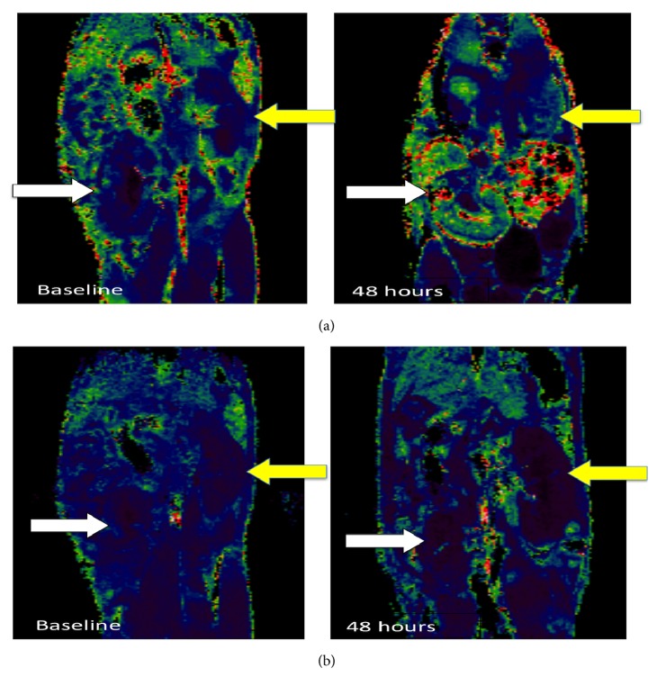
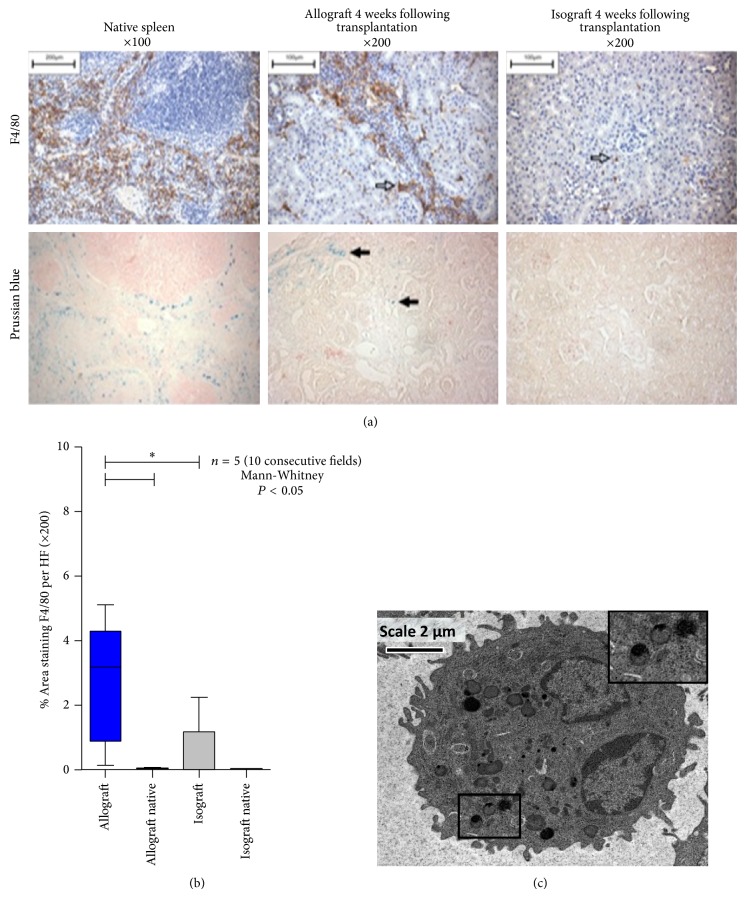
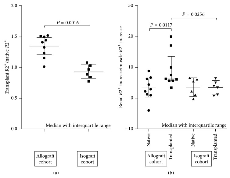
Objectives. We investigated whether ultrasmall paramagnetic particles of iron oxide- (USPIO-) enhanced magnetic resonance imaging (MRI) can detect experimental chronic allograft damage in a murine renal allograft model. Materials and Methods. Two cohorts of mice underwent renal transplantation with either a syngeneic isograft or allograft kidney. MRI scanning was performed prior to and 48 hours after USPIO infusion using T2(∗)-weighted protocols. R2(∗) values were calculated to indicate the degree of USPIO uptake. Native kidneys and skeletal muscle were imaged as reference tissues and renal explants analysed by histology and electron microscopy. Results. R2(∗) values in the allograft group were higher compared to the isograft group when indexed to native kidney (median 1.24 (interquartile range: 1.12 to 1.36) versus 0.96 (0.92 to 1.04), P < 0.01). R2(∗) values were also higher in the allograft transplant when indexed to skeletal muscle (6.24 (5.63 to 13.51)) compared to native kidney (2.91 (1.11 to 6.46) P < 0.05). Increased R2(∗) signal in kidney allograft was associated with macrophage and iron staining on histology. USPIO were identified within tissue resident macrophages on electron microscopy. Conclusion. USPIO-enhanced MRI identifies macrophage.
目的。我们研究了超小超顺磁性氧化铁颗粒(USPIO)增强磁共振成像(MRI)是否能够在小鼠肾移植模型中检测实验性慢性移植物损伤。材料与方法。两组小鼠分别接受同基因同系移植或同种异体移植肾移植。在注射USPIO之前和之后48小时,使用T2(∗)加权方案进行MRI扫描。计算R2(∗)值以指示USPIO摄取程度。将天然肾脏和骨骼肌作为参考组织进行成像,并通过组织学和电子显微镜分析肾外植体。结果。与同系移植组相比,同种异体移植组以天然肾脏为参照时的R2(∗)值更高(中位数1.24(四分位间距:1.12至1.36)对0.96(0.92至1.04),P<0.01)。与天然肾脏相比,同种异体移植以骨骼肌为参照时的R2(∗)值也更高(6.24(5.63至13.51))(2.91(1.11至6.46),P<0.05)。同种异体移植肾中R2(∗)信号增加与组织学上的巨噬细胞和铁染色相关。在电子显微镜下在组织驻留巨噬细胞内鉴定出USPIO。结论。USPIO增强MRI可识别巨噬细胞。